
Page 429 - Withrow and MacEwen's Small Animal Clinical Oncology, 6th Edition
P. 429
CHAPTER 22 Soft Tissue Sarcomas 407
In one study of 16 dogs with brachial plexus PNSTs treated with
limb-sparing compartmental resection, the overall MST was
1303 days and was significantly better for dogs with complete
VetBooks.ir histologic excision (MST 2227 days) compared with dogs with
51
incomplete excision (MST 487 days). For peripheral nerve
tumors extending through the foramen, hemilaminectomy may
be required in addition to forequarter amputation for adequate
tumor excision. Stereotactic RT has been described in 10 dogs
50
with brachial plexus tumors with partial or complete resolu-
tion of neurologic signs in all dogs. The mean progression-free
52
survival (PFS) and overall survival times (OSTs) were 240 days
and 371 days, respectively, with progression reported in 90% of
52
dogs. Regardless of histologic grade, local disease usually limits
survival before metastasis occurs. 50,52
Tumors of Adipose Tissue
Lipoma
Lipomas are benign tumors of adipose tissue, and can be differen-
tiated from liposarcomas based on morphologic, CT, and histo-
logic appearance. There are three morphologic types of lipomas:
53
regular, infiltrative, and intermuscular. 53–59 Histologically, lipo-
mas have indistinct nuclei and cytoplasm resembling normal fat,
whereas liposarcomas are characterized by increased cellularity,
distinct nuclei, and abundant cytoplasm with one or more drop-
lets of fat. Histologic variants of lipomas have been reported and
60
include angiolipoma and angiofibrolipoma. 61
Regular lipomas are relatively common in older dogs, espe-
cially in subcutaneous locations, and are rarely symptomatic.
They have been reported in the thoracic cavity, abdominal cav-
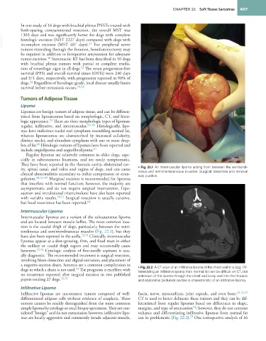
ity, spinal canal, and vulva and vagina of dogs, and can cause • Fig. 22.1 An intermuscular lipoma arising from between the semitendi-
clinical abnormalities secondary to either compression or stran- nosus and semimembranosus muscles. Surgical dissection and removal
gulation. 60,62–69 Marginal excision is recommended for lipomas was curative.
that interfere with normal function; however, the majority are
asymptomatic and do not require surgical intervention. Lipo-
suction and intralesional triamcinolone have also been reported
with variable results. 70,71 Surgical resection is usually curative,
68
but local recurrence has been reported.
Intermuscular Lipoma
Intermuscular lipomas are a variant of the subcutaneous lipoma
and are located between muscle bellies. The most common loca-
tion is the caudal thigh of dogs, particularly between the semi-
tendinosus and semimembranosus muscles (Fig. 22.1), but they
have also been reported in the axilla. 72,73 Clinically, intermuscular
lipomas appear as a slow-growing, firm, and fixed mass in either
the axillary or caudal thigh region and may occasionally cause
lameness. 72,73 Cytologic analysis of fine-needle aspirates is usu-
ally diagnostic. The recommended treatment is surgical resection,
involving blunt dissection and digital extrusion, and placement of
a negative-suction drain. Seromas are a common complication in
72
dogs in which a drain is not used. The prognosis is excellent with • Fig. 22.2 A CT scan of an infiltrative lipoma of the chest wall in a dog. Dif-
ferentiating an infiltrative lipoma from normal fat can be difficult on CT, but
no recurrence reported after surgical excision in two published extension of the lipoma through the chest and body wall into the thoracic
papers totaling 27 dogs. 72,73 and abdominal (pictured) cavities is characteristic of an infiltrative lipoma.
Infiltrative Lipoma
Infiltrative lipomas are uncommon tumors composed of well- fascia, nerve, myocardium, joint capsule, and even bone. 55,74,75
differentiated adipose cells without evidence of anaplasia. These CT is used to better delineate these tumors and they can be dif-
tumors cannot be readily distinguished from the more common ferentiated from regular lipomas based on differences in shape,
simple lipoma by cytology or small biopsy specimens. They are con- margins, and type of attenuation ; however, they do not contrast
53
sidered “benign” and do not metastasize; however, infiltrative lipo- enhance and differentiating infiltrative lipomas from normal fat
72
mas are locally aggressive and commonly invade adjacent muscle, can be problematic (Fig. 22.2). One retrospective analysis of 16