
Page 432 - Withrow and MacEwen's Small Animal Clinical Oncology, 6th Edition
P. 432
410 PART IV Specific Malignancies in the Small Animal Patient
The biopsy should be planned and positioned so that the biopsy
tract can be included in the curative-intent treatment with-
out increasing the surgical dose or size of the radiation field.
VetBooks.ir Although needle-core and incisional biopsies will typically
provide sufficient tissue for a definitive diagnosis of STS, the
determination of histologic grade from preoperative biopsies was
incorrect in 41% of dogs compared with the definitive surgical
sample, with histologic grade underestimated in 29% of dogs
and overestimated in 12% of dogs. 104 Excisional biopsies are not
recommended because they may not be curative and the subse-
quent surgery required to achieve complete histologic margins is
often more aggressive than surgery after core or incisional biop-
sies, resulting in additional morbidity and treatment costs. Fur-
thermore, multiple attempts at resection, including excisional
biopsy, before definitive therapy have a negative effect on ST in
dogs with STSs. 105
Diagnostic tests performed for workup and clinical stag-
ing include routine hematologic and serum biochemical blood
tests, three-view thoracic radiographs, abdominal ultrasonog-
raphy or advanced imaging, FNA or biopsy of the regional
LNs, and regional imaging of the STS. Three-view thoracic
radiographs should be performed before definitive treatment
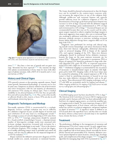
• Fig. 22.6 The typical gross appearance of a canine soft tissue sarcoma because the lungs are the most common metastatic site for
35
with a firm, well-circumscribed, expansile subcutaneous mass. typical STSs. Although LN metastasis is uncommon, FNA or
biopsy of regional LNs should be performed in dogs with clini-
cally abnormal LNs, grade III STSs, or suspected nonconven-
tissue. 92–101 They have a slow rate of growth and can grow very tional STSs with a high rate of metastasis to regional LNs (e.g.,
large. Metastasis has been reported. 96–100 The outcome for dogs HS). 106 Abdominal imaging is recommended for the assessment
with splenic mesenchymomas is better than for those with other of metastasis to intraabdominal organs in animals with high-
types of splenic sarcomas, with a MST of 12 months and a 1-year grade pelvic limb STS. Imaging studies of the local tumor may
96
survival rate of 50%. be required for planning of the surgical approach or RT if the
tumor is fixed to underlying structures or located in an area
History and Clinical Signs that may make definitive treatment difficult, such as the pel-
vic region. Three-dimensional (3D) imaging techniques such as
STSs generally present as slow-growing expansile masses. Rapid CT and MRI are particularly useful for staging local disease. 107
tumor growth, intratumoral hemorrhage, or necrosis can be seen Other imaging modalities for staging of the local tumor include
in some cases. Symptoms are directly related to site of involvement survey radiographs and ultrasonography. 108
and tumor invasiveness, with the vast majority of subcutaneous
and cutaneous STSs causing no clinical signs. There is marked Clinical Staging
variability in the physical features of STS, but they are generally
firm and well circumscribed (Fig. 22.6). They can be either mobile A modified staging system has been described for STSs in dogs.
26
or adherent (fixed) to skin, muscle, or bone. STSs can also be soft The American Joint Committee on Cancer (AJCC) staging system
and lobulated, mimicking lipomas. currently used in humans with STSs has been substantially modi-
fied from the original staging system, on which the modified ani-
Diagnostic Techniques and Workup mal staging system is based. The most important change to AJCC
staging is categorization of local disease, with less emphasis on
Fine-needle aspiration (FNA) is recommended for a cytologic tumor size, which is an arbitrary assignment, and greater emphasis
diagnosis; however, cytologic evaluation may not be sufficient on depth of invasion. 81,109 A superficial tumor is defined as an
for a definitive diagnosis because variable degrees of necrosis and STS located above the superficial fascia and that does not invade
poor exfoliation of cells may result in a nondiagnostic sample. the fascia, whereas a deep tumor is located deep to the superficial
26
The cytologic accuracy of correctly diagnosing an STS varies from fascia, invades the fascia, or both. 109
63% to 97%. 32,102 Cytologic preparations should be assessed
by a board-certified cytopathologist because a disproportionate Treatment
number of false-negative cytologic results were associated with
in-house cytologic assessments compared with evaluation by a The predominant challenge in the management of cutaneous and
33
board-certified cytopathologist in one study. Even in the absence subcutaneous STSs is local tumor control. As such, surgical resec-
of a definitive diagnosis, FNA cytology can exclude the diagno- tion is the principal treatment for dogs with STSs. RT may also
sis of readily exfoliating tumors such as epithelial and round cell play a significant role in local tumor control, especially for incom-
tumors, and this may be sufficient for the suspected diagnosis of pletely resected and unresectable STSs. However, definitive treat-
an STS by exclusion. 102,103 ment options depend on tumor location, clinical stage, histologic
Biopsy methods for definitive preoperative diagnosis of STSs grade, and completeness of histologic margins. 10,26,110 A suggested
include needle-core, punch, incisional, or excisional biopsies. algorithm for managing dogs with STSs is presented in Fig. 22.7.