
Page 430 - Withrow and MacEwen's Small Animal Clinical Oncology, 6th Edition
P. 430
408 PART IV Specific Malignancies in the Small Animal Patient
subtype was not prognostic, but metastatic disease was more com-
76
Surgery type mon in dogs with pleomorphic liposarcomas. A revised classifi-
cation scheme has been proposed on the basis of IHC expression
1.0
Marginal excision
VetBooks.ir 0.8 Incisional biopsy of MDM2 and CDK4. In one study, MDM2 and CDK4 were
Wide excision
expressed in 67% and 88% of well-differentiated liposarcomas,
Proportion surviving 0.6 phic liposarcomas, and 75% and 100% of dedifferentiated liposar-
14% and 71% of myxoid liposarcomas, 0% and 67% of pleomor-
84
comas. Furthermore, Ki67 index also correlated with histotype
and was lowest in well-differentiated liposarcomas and highest in
84
dedifferentiated liposarcomas. These results parallel the human
0.4
ated and dedifferentiated liposarcomas distinct entities, but that
0.2 data to some degree and suggest that not only are well-differenti-
84
this classification scheme may have prognostic significance.
0.0
0 1 2 3 4 5 Tumors of Skeletal Muscle
Days
Rhabdomyosarcoma
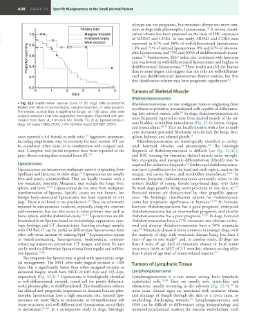
• Fig. 22.3 Kaplan–Meier survival curve of 56 dogs with liposarcoma Rhabdomyosarcomas are rare malignant tumors originating from
treated with either incisional biopsy, marginal resection, or wide excision. myoblasts or primitive mesenchymal cells capable of differentiat-
The median survival time is significantly longer, at 1188 days, after wide ing into striated muscle cells. In dogs, rhabdomyosarcomas are
85
surgical resection than less aggressive techniques. (Reprinted with per- most frequently reported to arise from skeletal muscle of the uri-
mission from Baez JL, Hendrick MJ, Shofer FS, et al: Liposarcomas in nary bladder, retrobulbar musculature (Fig. 22.4), larynx, tongue,
dogs: 56 cases (1989–2000), J Am Vet Med Assoc 224:887, 2004.)
and myocardium. 86,87 They are locally invasive with a low to mod-
erate metastatic potential. Metastatic sites include the lungs, liver,
57
cases reported a 4:1 female-to-male ratio. Aggressive treatment, spleen, kidneys, and adrenal glands. 85
including amputation, may be necessary for local control. RT can Rhabdomyosarcomas are histologically classified as embry-
be considered either alone or in combination with surgical exci- onal, botryoid, alveolar, and pleomorphic. The histologic
86
sion. Complete and partial responses have been reported in the diagnosis of rhabdomyosarcoma is difficult (see Fig. 22.4C),
59
gross disease setting after external beam RT. and IHC staining for vimentin, skeletal muscle actin, myoglo-
bin, myogenin, and myogenic differentiation (MyoD) may be
Liposarcoma required for definitive diagnosis. Embryonal rhabdomyosarco-
88
Liposarcomas are uncommon malignant tumors originating from mas have a predilection for the head and neck region, such as the
lipoblasts and lipocytes in older dogs. Liposarcomas are usually tongue, oral cavity, larynx, and retrobulbar musculature. 86,87 In
76
firm and poorly circumscribed. They are locally invasive with a contrast, botryoid rhabdomyosarcoma commonly arises in the
low metastatic potential. Metastatic sites include the lungs, liver, urinary bladder of young, female large-breed dogs, with Saint
spleen, and bone. 29,76 Liposarcomas do not arise from malignant Bernard dogs possibly being overrepresented in one data set.
85
transformation of lipomas. Specific causes are not known, but Botryoid tumors are characterized by their grapelike appear-
foreign body–associated liposarcoma has been reported in one ance. The histologic classification scheme for rhabdomyosar-
76
dog. There is no breed or sex predilection. They are commonly coma has prognostic significance in humans. 85,89 In humans,
5
reported in subcutaneous locations, especially along the ventrum botryoid rhabdomyosarcoma has a good prognosis, embryonal
and extremities, but can also occur in other primary sites such as rhabdomyosarcoma has an intermediate prognosis, and alveolar
bone, spleen, and the abdominal cavity. 76–78 Liposarcomas are dif- rhabdomyosarcoma has a poor prognosis. 85,89 In dogs, botryoid
ferentiated from lipomas based on morphologic appearance, cyto- rhabdomyosarcomas have a 27% metastatic rate whereas embry-
logic findings, and CT characteristics. Staining cytologic samples onal and alveolar rhabdomyosarcomas have a 50% metastatic
with Oil Red O can be useful to differentiate liposarcomas from rate. Metastatic disease is more common in younger dogs, with
86
other soft tissue saromas by staining lipid. Liposarcomas appear the majority of dogs with metastatic disease being less than 2
79
as mixed-attenuating, heterogeneous, multinodular, contrast- years of age in one study and, in another study, all dogs less
86
enhancing masses on precontrast CT images, and these features than 4 years of age died of metastatic disease or local tumor
can be used to differentiate liposarcomas from regular and infiltra- recurrence (with an MST of 2.5 months), whereas no dog older
87
tive lipomas. 53,80 than 4 years of age died of tumor-related reasons.
The prognosis for liposarcoma is good with appropriate surgi-
cal management. The MST after wide surgical excision is 1188 Tumors of Lymphatic Tissue
days; this is significantly better than either marginal excision or
incisional biopsy, which have MSTs of 649 days and 183 days, Lymphangiosarcoma
76
respectively (Fig. 22.3). Liposarcoma is histologically classified Lymphangiosarcoma is a rare tumor arising from lymphatic
as well-differentiated, myxoid, round cell (or poorly differenti- endothelial cells. 29,90 They are usually soft, cystic-like, and
29
ated), pleomorphic, or dedifferentiated. This classification scheme edematous, usually occurring in the subcutis (Fig. 22.5). In
has clinical and prognostic importance in humans because pleo- most cases, clinical signs are associated with extensive edema
morphic liposarcomas have a high metastatic rate, myxoid lipo- and drainage of lymph through the skin or a cystic mass, or
sarcomas are more likely to metastasize to extrapulmonary soft nonhealing, discharging wounds. Lymphangiosarcoma and
90
tissue structures, and well-differentiated liposarcomas are unlikely HSA can be difficult to differentiate using histopathology and
to metastasize. 81–83 In a retrospective study in dogs, histologic immunohistochemical markers for vascular endothelium, such