
Page 431 - Withrow and MacEwen's Small Animal Clinical Oncology, 6th Edition
P. 431
CHAPTER 22 Soft Tissue Sarcomas 409
VetBooks.ir
B
A
C
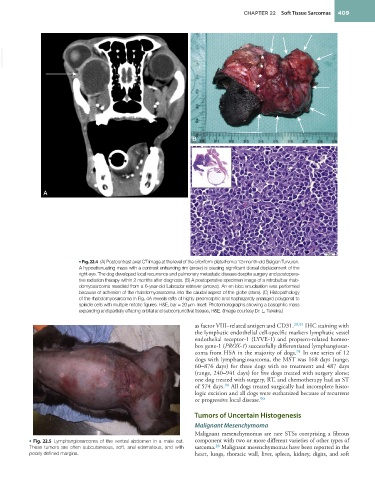
• Fig. 22.4 (A) Postcontrast axial CT image at the level of the cribriform plate from a 12-month-old Belgian Turvuren.
A hypoattenuating mass with a contrast enhancing rim (arrow) is causing significant dorsal displacement of the
right eye. The dog developed local recurrence and pulmonary metastatic disease despite surgery and postopera-
tive radiation therapy within 2 months after diagnosis. (B) A postoperative specimen image of a retrobulbar rhab-
domyosarcoma resected from a 6-year-old Labrador retriever (arrows). An en bloc enucleation was performed
because of adhesion of the rhabdomyosarcoma into the caudal aspect of the globe (stars). (C) Histopathology
of the rhabdomyosarcoma in Fig. 4A reveals rafts of highly pleomorphic and haphazardly arranged polygonal to
spindle cells with multiple mitotic figures. H&E, bar = 20 μm. Inset: Photomicrographs showing a basophilic mass
expanding and partially effacing orbital and subconjunctival tissues, H&E. (Image courtesy Dr. L. Teixeira.)
as factor VIII–related antigen and CD31. 29,91 IHC staining with
the lymphatic endothelial cell-specific markers lymphatic vessel
endothelial receptor-1 (LYVE-1) and propsero-related homeo-
box gene-1 (PROX-1) successfully differentiated lymphangiosar-
coma from HSA in the majority of dogs. In one series of 12
91
dogs with lymphangiosarcoma, the MST was 168 days (range,
60–876 days) for three dogs with no treatment and 487 days
(range, 240–941 days) for five dogs treated with surgery alone;
one dog treated with surgery, RT, and chemotherapy had an ST
90
of 574 days. All dogs treated surgically had incomplete histo-
logic excision and all dogs were euthanized because of recurrent
90
or progressive local disease.
Tumors of Uncertain Histogenesis
Malignant Mesenchymoma
Malignant mesenchymomas are rare STSs comprising a fibrous
• Fig. 22.5 Lymphangiosarcoma of the ventral abdomen in a male cat. component with two or more different varieties of other types of
26
These tumors are often subcutaneous, soft, and edematous, and with sarcoma. Malignant mesenchymomas have been reported in the
poorly defined margins. heart, lungs, thoracic wall, liver, spleen, kidney, digits, and soft