
Page 436 - Withrow and MacEwen's Small Animal Clinical Oncology, 6th Edition
P. 436
VetBooks.ir
A B
C D
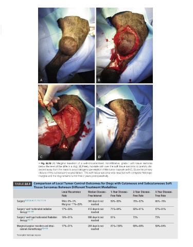
• Fig. 22.10 (A) Marginal resection of a well-circumscribed, noninfiltrative, grade I soft tissue sarcoma
below the level of the stifle in a dog. (B) Freely movable skin over the soft tissue sarcoma is carefully dis-
sected away from the mass to avoid iatrogenic penetration of the tumor capsule and (C, D) permit primary
closure of the subsequent wound defect. This soft tissue sarcoma was resected with complete histologic
margins and the dog remains tumor-free 2 years postoperatively.
TABLE 22.3 Comparison of Local Tumor Control Outcomes for Dogs with Cutaneous and Subcutaneous Soft
Tissue Sarcomas Between Different Treatment Modalities
Local Recurrence Median Disease- 1-Year Disease- 2-Year Disease- 3-Year Disease-
Rate Free Interval Free Rate Free Rate Free Rate
Surgery 25,32,34,46,111,113,117,118 Wide: 0%–5% 368 days to not 89%–93% 78%–82% 66%–76%
Marginal: 11%–29% reached
a
Surgery and fractionated radiation 17%–39% 412 days to not 71%–84% 60%–81% 57%–81%
therapy 135–138 reached
a
Surgery and hypofractionated Radiation 18%–21% 698 days to not 81% 73% 73%
therapy 140,141 reached
Marginal surgical resection and intral- 17%–31% 264 days to not 81%–100% 69%–89% 59%–84%
esional chemotherapy 162–164 reached
a Incomplete histologic excision.