
Page 441 - Withrow and MacEwen's Small Animal Clinical Oncology, 6th Edition
P. 441
CHAPTER 22 Soft Tissue Sarcomas 419
VetBooks.ir
A
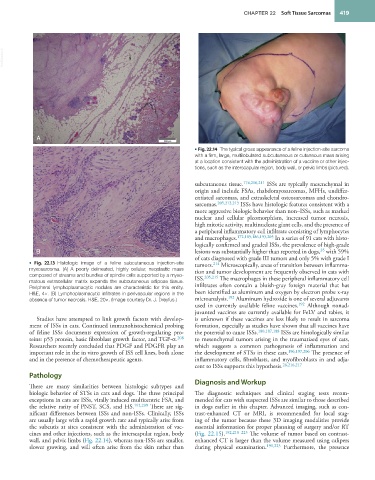
• Fig. 22.14 The typical gross appearance of a feline injection-site sarcoma
with a firm, large, multilobulated subcutaneous or cutaneous mass arising
at a location consistent with the administration of a vaccine or other injec-
tions, such as the interscapular region, body wall, or pelvic limbs (pictured).
subcutaneous tissue. 174,210,211 ISSs are typically mesenchymal in
origin and include FSAs, rhabdomyosarcomas, MFHs, undiffer-
entiated sarcomas, and extraskeletal osteosarcomas and chondro-
sarcomas. 205,212,213 ISSs have histologic features consistent with a
more aggressive biologic behavior than non–ISSs, such as marked
nuclear and cellular pleomorphism, increased tumor necrosis,
high mitotic activity, multinucleate giant cells, and the presence of
a peripheral inflammatory cell infiltrate consisting of lymphocytes
and macrophages. 172,185,186,193,205 In a series of 91 cats with histo-
logically confirmed and graded ISSs, the prevalence of high-grade
35
B lesions was substantially higher than reported in dogs, with 59%
of cats diagnosed with grade III tumors and only 5% with grade I
• Fig. 22.13 Histologic image of a feline subcutaneous injection-site tumors. 214 Microscopically, areas of transition between inflamma-
myxosarcoma. (A) A poorly delineated, highly cellular, neoplastic mass tion and tumor development are frequently observed in cats with
composed of streams and bundles of spindle cells supported by a myxo- ISS. 205,215 The macrophages in these peripheral inflammatory cell
matous extracellular matrix expands the subcutaneous adipose tissue. infiltrates often contain a bluish-gray foreign material that has
Peripheral lymphoplasmacytic nodules are characteristic for this entity.
H&E, 4×. (B) Lymphoplasmacytic infiltrates in perivascular regions in the been identified as aluminum and oxygen by electron probe x-ray
absence of tumor necrosis. H&E, 20×. (Image courtesy Dr. J. Dreyfus.) microanalysis. 192 Aluminum hydroxide is one of several adjuvants
used in currently available feline vaccines. 192 Although nonad-
juvanted vaccines are currently available for FeLV and rabies, it
Studies have attempted to link growth factors with develop- is unknown if these vaccines are less likely to result in sarcoma
ment of ISSs in cats. Continued immunohistochemical probing formation, especially as studies have shown that all vaccines have
of feline ISSs documents expression of growth-regulating pro- the potential to cause ISSs. 180,187,188 ISSs are histologically similar
teins: p53 protein, basic fibroblast growth factor, and TGF-α. 208 to mesenchymal tumors arising in the traumatized eyes of cats,
Researchers recently concluded that PDGF and PDGFR play an which suggests a common pathogenesis of inflammation and
important role in the in vitro growth of ISS cell lines, both alone the development of STSs in these cats. 196,197,206 The presence of
and in the presence of chemotherapeutic agents. inflammatory cells, fibroblasts, and myofibroblasts in and adja-
cent to ISSs supports this hypothesis. 26,216,217
Pathology
Diagnosis and Workup
There are many similarities between histologic subtypes and
biologic behavior of STSs in cats and dogs. The three principal The diagnostic techniques and clinical staging tests recom-
exceptions in cats are ISSs, virally induced multicentric FSA, and mended for cats with suspected ISSs are similar to those described
the relative rarity of PNST, SCS, and HS. 192,209 There are sig- in dogs earlier in this chapter. Advanced imaging, such as con-
nificant differences between ISSs and non-ISSs. Clinically, ISSs trast-enhanced CT or MRI, is recommended for local stag-
are usually large with a rapid growth rate and typically arise from ing of the tumor because these 3D imaging modalities provide
the subcutis at sites consistent with the administration of vac- essential information for proper planning of surgery and/or RT
cines and other injections, such as the interscapular region, body (Fig. 22.15). 192,218–223 The volume of tumor based on contrast-
wall, and pelvic limbs (Fig. 22.14), whereas non-ISSs are smaller, enhanced CT is larger than the volume measured using calipers
slower growing, and will often arise from the skin rather than during physical examination. 191,223 Furthermore, the presence