
Page 442 - Withrow and MacEwen's Small Animal Clinical Oncology, 6th Edition
P. 442
420 PART IV Specific Malignancies in the Small Animal Patient
for wide resection or radical surgery. 224 In addition, the median
time to first recurrence is only 66 days when the first surgery is
performed at a nonreferral institution compared with 274 days
VetBooks.ir at referral institutions. 224 Inadequate biopsy planning, preop-
erative staging, and/or attempts at first surgery will result in an
increase in tumor margins and may make further surgical treat-
ment more difficult to impossible. The first attempt at surgical
management of cats with ISSs should be performed by a refer-
R L
ral surgeon with experience in aggressive resection, especially in
the interscapular, body wall, or pelvic regions, to increase the
chance of a successful outcome. 214,224–228 Similar to dogs with
STSs, biopsy tracts and any areas of fixation, including bone and
fascia, should be resected en bloc with the tumor. In cats with
ISSs, wide surgical resection of tumors located in the interscapu-
lar region will often involve excision of dorsal spinous processes
(Fig. 22.16), whereas thoracic or body wall resection is often
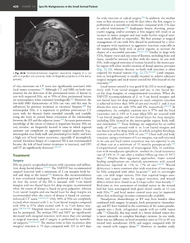
• Fig. 22.15 Contrast-enhanced magnetic resonance imaging of a cat required for truncal tumors (Fig. 22.17). 224,225 Limb amputa-
with an injection-site sarcoma. Note the fingerlike projections of the tumor tion or hemipelvectomy is usually required to achieve adequate
(arrows). surgical margins and local tumor control for ISSs located on the
extremity. 214
of skip metastases on CT scans was significantly associated with Local tumor control is very good if the ISS is treated aggres-
local tumor recurrence. 222 Although CT and MRI are both very sively with 5-cm lateral margins and one to two fascial lay-
sensitive for the detection of the peritumoral extent of disease in ers for deep margins, or compartmental resections. When the
cats with suspected ISSs, up to 59% of these peritumoral lesions VAFSTF recommendations of 2- to 3-cm lateral margins and
are nonneoplastic when examined histologically. 221 Moreover, the one fascial layer for deep margins are used, complete resection
low-field MRI characteristics of ISSs can vary and this may be is achieved in fewer than 50% of cats and overall 1- and 2-year
influenced by previous incisional or excisional biopsies. 218 For disease-free rates are only 35% and 9%, respectively. 224,225 In
interscapular ISSs, it is important to perform postcontrast CT comparison, the complete excision rate is 97% and the local
scans with the thoracic limbs extended cranially and caudally recurrence rate is 14% at 3 years when ISSs are resected with
along the body to permit better evaluation of the relationship 5-cm lateral margins and two fascial layers for deep margins,
between the ISS and the adjacent tissue. 219 Accurate pretreatment including ISSs located in the interscapular region, body wall,
knowledge of the extent of disease is important because ISSs are and extremities. 214 These findings are supported by an earlier
very invasive, are frequently located in areas in which regional study of 57 cats treated with 4- to 5-cm lateral margins and
anatomy can complicate an aggressive surgical approach (e.g., one fascial layer for deep margins, in which complete histologic
interscapular area, body wall, and proximal pelvic limb), and have excision was achieved in 95% of cats. 226 Chest wall and body
a high rate of local tumor recurrence, especially if incompletely resection, using a minimum of 3-cm margins, was well tolerated
excised. Excisional biopsy of a suspected ISS is not recommended in six cats and local tumor recurrence was not reported in any
because the risk of local tumor recurrence is increased, and DFI of these cats at a minimum of 12 months postoperatively. 228
and ST are significantly decreased. 224,225 Compartmental resections of interscapular ISSs, in combina-
tion with neoadjuvant epirubicin, resulted in a local recurrence
Treatment rate of 14% in 21 cats after a median follow-up time of 1072
Despite these aggressive approaches, major wound
days.
229
Surgery healing complications are relatively uncommon, with wound
ISSs are poorly encapsulated tumors with extension and infiltra- dehiscence reported in 11% to 17% of cats. 214,227 Wound
tion along fascial planes. 215,216 The VAFSTF has recommended dehiscence is more common after wide resection of interscapu-
surgical resection with a minimum of 2 cm margins both lat- lar ISSs compared with other locations 214 and in overweight
eral and deep to the tumor 192 ; however, this recommendation cats, cats with larger tumors, ISSs that required longer anes-
is now considered inadequate. The preferred approach is based thetic and surgical times for surgical resection, and wound
on how the extent of the ISS is assessed, with 5-cm lateral defects which were closed in an X-shape rather than linearly. 227
margins and two fascial layers for deep margins recommended Real-time in vivo assessment of residual tumor in the wound
when the extent of disease is based on gross palpation, whereas bed has been investigated with good initial results in 12 cats
3-cm lateral margins and one fascial layer for deep margins are with ISSs, 230 and local tumor control rates may be improved
recommended when the extent of disease is based on contrast- when this technology becomes commercially available.
enhanced CT scans. 214,226,227 Only 50% of ISSs are completely Neoadjuvant chemotherapy or RT may have benefits when
excised when resected with 2- to 3-cm lateral margins compared combined with surgery. In people, both preoperative chemother-
with 95% to 97% when excised with 4- to 5-cm lateral mar- apy and RT have resulted in the conversion of the tumor pseudo-
gins. 214,224–226 Marginal resection or excisional biopsy should capsule into a thick, collagenized capsule with no viable tumor
not be attempted. The median DFI and MST are significantly cells. 114 Clinically, this may result in a better defined tumor that
decreased with marginal resection, with more than one attempt is more amenable to complete histologic excision. In one study,
at surgical resection, and if surgery is performed by nonrefer- there was no difference in local recurrence rates or STs in 49 cats
ral surgeons. 214,224,225 The median time to first recurrence after treated with neoadjuvant DOX and surgery compared with 20
marginal resection is 79 days compared with 325 to 419 days cats treated with surgery alone. 231 However, in another study,