
Page 572 - Withrow and MacEwen's Small Animal Clinical Oncology, 6th Edition
P. 572
550 PART IV Specific Malignancies in the Small Animal Patient
VetBooks.ir
A B
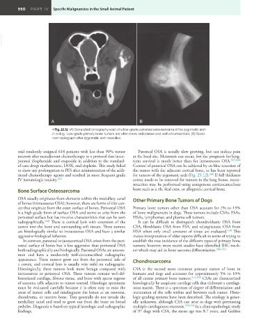
• Fig. 25.12 (A) Computed tomography scan of a low-grade parosteal osteosarcoma of the zygomatic arch
in a dog. Low-grade primary bone tumors are often more radiodense and well circumscribed. (B) Speci-
men radiograph after zygomatic arch resection.
trial randomly assigned 618 patients with less than 90% tumor Parosteal OSA is usually slow growing, but can induce pain
necrosis after neoadjuvant chemotherapy to a protocol that incor- at the local site. Metastasis can occur, but the prognosis for long-
porated ifosphamide and etoposide in addition to the standard- term survival is much better than for intraosseous OSA. 347,348
of-care drugs methotrexate, DOX, and cisplatin. This study failed Control of parosteal OSA can be achieved by en bloc resection of
to show any prolongation in PFS after administration of the addi- the tumor with the adjacent cortical bone, as has been reported
tional chemotherapy agents and resulted in more frequent grade for tumors of the zygomatic arch (Fig. 25.12). 348 If full thickness
IV hematologic toxicity. 345 cortex needs to be removed for tumors in the long bones, recon-
struction may be performed using autogenous corticocancellous
Bone Surface Osteosarcoma bone such as a rib, ileal crest, or allogeneic cortical bone.
OSA usually originates from elements within the medullary canal Other Primary Bone Tumors of Dogs
of bones (intraosseous OSA); however, there are forms of this can-
cer that originate from the outer surface of bones. Periosteal OSA Primary bone tumors other than OSA account for 2% to 15%
is a high-grade form of surface OSA and seems to arise from the of bone malignancies in dogs. These tumors include CSAs, FSAs,
periosteal surface but has invasive characteristics that can be seen HSAs, lymphomas, and plasma cell tumors.
radiographically. 346 There is cortical lysis with extension of the It can be difficult to distinguish chondroblastic OSA from
tumor into the bone and surrounding soft tissues. These tumors CSA, fibroblastic OSA from FSA, and telangiectatic OSA from
are histologically similar to intraosseous OSA and have a similar HSA when only small amounts of tissue are evaluated. 140 This
aggressive biological behavior. makes interpretation of older reports difficult in terms of trying to
In contrast, parosteal or juxtacortical OSA arises from the peri- establish the true incidence of the different types of primary bone
osteal surface of bones but is less aggressive than periosteal OSA tumors; however, more recent studies have identified IHC mark-
both radiographically and biologically. Parosteal OSAs are uncom- ers which may aid in bone sarcoma differentiation. 349–351
mon and have a moderately well-circumscribed radiographic
appearance. These tumors grow out from the periosteal side of Chondrosarcoma
a cortex, and cortical lysis is usually very mild on radiographs.
Histologically, these tumors look more benign compared with CSA is the second most common primary tumor of bone in
intraosseous or periosteal OSA. These tumors contain well-dif- humans and dogs and accounts for approximately 5% to 10%
ferentiated cartilage, fibrous tissue, and bone with sparse regions of all canine primary bone tumors. 2–5,352 CSAs are characterized
of sarcoma cells adjacent to tumor osteoid. Histologic specimens histologically by anaplastic cartilage cells that elaborate a cartilagi-
must be evaluated carefully because it is often easy to miss the nous matrix. There is a spectrum of degree of differentiation and
areas of tumor cells and misdiagnose the lesion as an osteoma, maturation of the cells within and between each tumor. Histo-
chondroma, or reactive bone. They generally do not invade the logic grading systems have been described. The etiology is gener-
medullary canal and tend to grow out from the bone on broad ally unknown, although CSA can arise in dogs with preexisting
pedicles. Diagnosis is based on typical histologic and radiographic multiple cartilaginous exostosis. 353,354 In a clinicopathologic study
findings. of 97 dogs with CSA, the mean age was 8.7 years, and Golden