
Page 573 - Withrow and MacEwen's Small Animal Clinical Oncology, 6th Edition
P. 573
CHAPTER 25 Tumors of the Skeletal System 551
Retrievers were at a higher risk of developing CSA than any other
breed. 355 There was no sex predilection, and 61% of the tumors
occurred on flat bones. CSA can originate in the nasal cavity, ribs,
VetBooks.ir long bones, pelvis, extraskeletal sites (such as the mammary gland,
heart valves, aorta, larynx, trachea, lung, and omentum), verte-
brae, facial bones, digits, and os penis. 29,355–362 The nasal cavity is
the most common site for canine CSA. 355
CSA is generally considered to have a lower metastatic rate
than OSA; however, a more aggressive variant, dedifferentiated
CSA, has been described in seven dogs and one cat, and the
metastatic rate in these animals was 63%. 363 Tumor location
rather than histologic grade was prognostic in one study, 344
but histologic grade was prognostic in two other studies. 364,365
The MST for dogs with nasal CSA ranges from 210 days to
580 days with various treatments (RT, rhinotomy and RT,
and rhinotomy alone). 355,366 Metastatic disease is rare in dogs
with nasal CSA. The MST for dogs with CSA of ribs var-
ies widely. 20,171,367 Reports before 1992 contained few cases
that were treated with intent to cure, but MSTs in more con-
temporary reports range from 1080 days to more than 3820
days. 172,181,364,365 The overall MST for 25 dogs with appen-
dicular CSA treated with limb amputation alone was 979
days, but outcomes were dependent on histologic grade. The
metastatic rates and MSTs for grade I, II, and III appendicular
CSAs were 0% and 6.0 years, 31% and 2.7 years, and 50% and
0.9 years, respectively. A reliable adjuvant chemotherapeutic
agent is not known for canine CSA.
Hemangiosarcoma
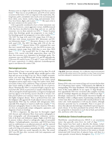
Primary HSA of bone is rare and accounts for less than 5% of all • Fig. 25.13 Specimen radiograph of a multilobular osteochondrosarcoma
bone tumors. This disease generally affects middle-aged to older arising from the vertical ramus of the mandible in a dog. These tumors have
dogs and can occur in dogs of any size. This is a highly metastatic a granular radiographic appearance often referred to as “popcorn ball.”
tumor, and most dogs affected will develop metastatic disease
within 6 months of diagnosis. Metastases can be widely spread Fibrosarcoma
throughout various organs such as lungs, liver, spleen, heart, skele-
tal muscles, kidney, brain, and bones. Dogs can present with mul- Primary FSA is also a rare tumor of dogs and accounts for less than
4
tiple lesions making it difficult to determine the site of primary 5% of all primary bone tumors. Unfortunately, the difficulty in
disease. Histologically, HSA is composed of highly anaplastic mes- distinguishing FSA from fibroblastic OSA histologically renders
enchymal cells, which are precursors to vascular endothelium. The study of this tumor difficult. In one report, 11 dogs thought to
cells are arranged in chords separated by a collagenous background have appendicular FSA were reevaluated after complete resection
and may appear to be forming vascular channels or sinuses. Cel- and the histologic diagnosis was changed to OSA in six dogs. 369
lular pleomorphism and numerous mitotic figures are features of Histologic characteristics of FSA include interwoven bundles of
this highly malignant disease. There is profound bone lysis, and fibroblasts within a collagen matrix of permeating cancellous and
the malignant cells aggressively invade adjacent normal structures. cortical bone that is not associated with osteoid produced by the
Appendicular HSA may be confused with telangiectatic OSA, tumor cells. Limb amputation or LSS may be curative, although
especially if the diagnosis is based on small tissue samples. 349 The metastatic potential may be considerable. There is no good evi-
dominant radiographic feature is often lysis; however, HSA does dence that adjuvant chemotherapy is beneficial in preventing met-
not have an unequivocally unique radiographic appearance, and astatic disease. It has been postulated that primary FSA of bone
diagnosis is based on histology. has a propensity to metastasize to such sites as heart, pericardium,
If HSA is diagnosed, the dog must be thoroughly staged skin, and bones rather than lung. 369
with thoracic and abdominal films, bone survey radiographs or
bone scintigraphy, and ultrasonographic evaluation, particu- Multilobular Osteochondrosarcoma
larly of the heart and abdominal organs. Right atrial HSA may
be present without clinical or radiographic signs of pericardial Multilobular osteochondrosarcoma (MLO) is an uncommon
effusion. Cyclophosphamide, vincristine, and DOX have been tumor that generally arises from the skull of dogs. 162,370–372
used in combination as an adjuvant protocol, and the reported Many names have been used to describe this disease, includ-
MST for dogs with nonskeletal HSA is 172 days. 368 In a recent ing chondroma rodens, multilobular osteoma, and multilobular
study of 41 dogs with primary appendicular HSA, a predilection tumor of bone. These tumors have a characteristic appearance on
for the pelvic limb was noted, especially the tibia. The overall radiographs, CT, and MRI; the borders of the tumor are sharply
MST was 299 days after treatment with limb amputation and demarcated with limited lysis of adjacent bone, and there is a
chemotherapy. 350 coarse granular mineral density throughout (Fig. 25.13). 373,374