
Page 629 - Withrow and MacEwen's Small Animal Clinical Oncology, 6th Edition
P. 629
CHAPTER 28 Tumors of the Mammary Gland 607
from molecular classification to inform precise and patient-spe- disease, 105,106 incorporation of human LN staging techniques
cific treatment recommendations in canine MGTs, similar to the should be reconsidered for the dog. 107 Sentinel LN mapping is a
means of detecting which LNs are receiving draining tumor lymph
approach in human BC therapy.
VetBooks.ir and thus most at risk for metastasis. Some techniques described
for sentinel LN mapping in the dog include lymphoscintigraphy
History and Clinical Presentation
using technetium, contrast-enhanced ultrasonography, autog-
MGTs are usually easy to detect through routine physical exami- enous hemosiderin, computed tomographic indirect lymphogra-
nations; however, high-risk dogs, specifically older intact female phy, and intraoperative dyes (Fig. 28.2). 103,104,107–111 Clinically
dogs, should undergo a thorough examination of the MGs. MGTs normal LNs can be difficult to identify in dogs with MGTs, as
typically affect the two caudal glands where the MGs or tissues the axillary LNs are often not palpable and the superficial inguinal
are naturally larger; thus careful palpation may be necessary to LNs reside deep to the fifth MG in the inguinal fat pad. Further-
detect small tumors. 9,14,66,67 The MGs should be palpated again more, many dogs have more than one tumor; thus multiple LNs
under general anesthesia to ensure that all tumors are found and may represent draining LNs. Lymphatic drainage of normal MGs
included in the surgical planning, and both chains should be care- is in addition very complex with documented drainage occurring
fully evaluated. A recent study documented that 70% of intact to multiple ipsilateral LNs and even to contralateral LNs. 104,112,113
15
females had more than one MGT at diagnosis. The size of the The amount of variation in lymphatic drainage increases in the
tumor(s), stage of disease, and presence of systemic signs of illness neoplastic MG. 112 Tumor-induced lymphangiogenesis, well docu-
vary widely. Inflammatory mammary carcinomas represent a rare mented in human BC, may be responsible for the unpredictable
but clinically important subset of MGTs in dogs. Affected dogs and erratic location of susceptible or “at-risk” LNs in dogs having
may easily be misdiagnosed as having mastitis or severe dermatitis malignant MGTs. 114 Thus exclusive anatomic sampling of nearby
because, rather than presenting with discrete well-circumscribed LNs may not be sufficient for accurate LN staging and may miss
tumors, the entire mammary chain may appear edematous, swol- the presence of locoregional disease. 107
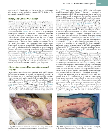
len, warm, and painful (Fig. 28.1). 101,102 In addition to the exten- The first description of sentinel LN mapping in any animal was
sive locoregional involvement, most dogs with inflammatory done in cats almost 30 years ago. 115 Benefits in human medicine
carcinomas have distant metastatic disease and signs of systemic include greater ease in identifying the at-risk LNs intraoperatively
illness. 101,102 These dogs are therefore poor surgical candidates. with minimal surgical incisions and with efficiency, especially for
The majority of dogs with MGTs are systemically healthy and the the rarely assessed axillary nodes. The prognostic value of sentinel
tumors are confined to the MGs when they are diagnosed. LN mapping is currently unknown for the dog, 116 although a study
of computed tomographic indirect lymphography found imaging
Clinical Assessment, Diagnosis, Workup, and characteristics of sentinel LNs positive for histologic mammary
tumor metastasis having decreased or heterogeneous LN contrast
Staging enhancement compared with the homogeneous contrast enhance-
Because of the risk of metastasis associated with MGTs, staging ment pattern of sentinel LNs negative for histologic metastasis. 103
before initiating therapy is strongly recommended, especially if Abdominal ultrasound may be indicated in dogs with sus-
benign disease cannot be histologically confirmed. Minimal diag- pected regional LN involvement or changes on preoperative
nostic workup and staging can include complete blood count blood work suggesting tumor-related or non–tumor-related
(CBC), serum biochemistry, three-view thoracic radiographs, and serum biochemistry changes. Even though osseous metaplasia
fine-needle aspiration (FNA) of regional lymph nodes (LNs) or occurs occasionally with mammary adenocarcinoma and the
more accurately, sentinel LN mapping and biopsy (see Chapter MGs are a common site for extraskeletal osteosarcoma (OSA),
9). 103,104 In women, the use of sentinel LN mapping has dramati- there has been no prognostic value found for serum alkaline
cally altered the surgical treatment of BC. As canine mammary phosphatase activity. 117,118 There may be value in perform-
carcinoma has been demonstrated as a relevant model for human ing MGT cytology to help rule out nonmammary dermal
• Fig. 28.1 Inflammatory mammary carcinoma in a dog. (Courtesy Dr. • Fig. 28.2 Regional mastectomy in a dog. (Courtesy Dr. Julius Liptak,
Nicholas Bacon, Fitzpatrick Referrals, Guildford, United Kingdom.) VCA Canada—Alta Vista Animal Hospital, Ottawa, Canada.)