
Page 632 - Withrow and MacEwen's Small Animal Clinical Oncology, 6th Edition
P. 632
610 PART IV Specific Malignancies in the Small Animal Patient
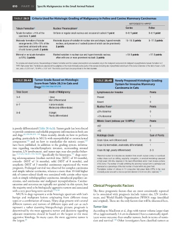
TABLE 28.3 Criteria Used for Histologic Grading of Malignancy in Feline and Canine Mammary Carcinomas
b
MITOSES/10 HPFS
VetBooks.ir Tubule Formation a Nuclear Pleomorphism a Canine Feline
Tubule formation >75% of the
specimen: 1 point Uniform or regular small nucleus and occasional nucleoli: 1 point 0–9: 1 point 0–8: 1 point
Moderate formation of tubular Moderate degree of variation in nuclear size and shape, hyperchromatic 10–19: 2 points 9–17: 2 points
arrangements (10%–75% of the nucleus, and presence of nucleoli (some of which can be prominent):
specimen) admixed with areas 2 points
of solid tumor growth: 2 points
Minimal or no tubule formation Marked variation in nuclear size and hyperchromatic nucleus, >19: 3 points >17: 3 points
(<10%): 3 points often with one or more prominent nucleoli: 3 points
a In complex and mixed tumors, the percentage of tubular formation and the nuclear pleomorphism are evaluated only in the malignant component. In malignant myoepithelioma tubular formation is 2
b HPF, High-power field. The fields are selected at the periphery or the most mitotically active parts of the sample (including also myeopithelial cells if part of the tumor). Diameter of the field of view = 0.55
2
2
mm; area = 0.237 mm ; 10 HPFs = 2.37 mm . The mitotic count has to be proportional to the field diameter.
TABLE 28.4A Tumor Grade Based on Histologic TABLE 28.4B Newly Proposed Histologic Grading
Score from Table 28.3 in Cats and System for Invasive Mammary
Dogs 133,134,136,137,210 Carcinoma in Cats
Total Score Grade of Malignancy Lymphovascular Invasion Points
3–5 I (low) Present 1
Well differentiated
Absent 0
6–7 II (intermediate) a
Moderately differentiated Nuclear Form Points
8–9 III (high) ≤5% Abnormal 0
Poorly differentiated >5% Abnormal 1
Mitotic Count (mitoses per 10 HPFs) b Points
≤62 0
is poorly differentiated (Table 28.4a,b). Tumor grade has been found
to provide consistent and reliable prognostic information in both cats >62 1
and dogs. 82,129,130,132–135 More recently, details on how to perform Histologic Grade Sum of Points
grading, particularly in MGTs with myoepithelial or mesenchymal
components 136 and on how to standardize the mitotic count, 137 Grade I (low, well differentiated) 0
have been published. In addition to the grading system, informa- Grade II (intermediate, moderately differentiated) 1
tion regarding vascular/lymphatic invasion, surrounding stromal
invasion, LN involvement, and tumor type may also predict behav- Grade III (high, poorly differentiated) 2–3
ior. 13,29,65,130,131,135,138,139 Specifically for histotypes, 131 dogs carry- a Abnormal nuclear form includes any deviation from smooth nuclear contour or round/oval
ing adenosquamous (median survival time [MST] of 18 months), nuclear shape such as clefting, angularity, corrugation, or ameboid morphology assessed
comedo- (MST of 14 months), solid (MST of 8 months), and at high power (40–60× objective) in the least differentiated and/or most invasive portions
anaplastic (MST of 3 months) carcinomas experienced the worst of the tumor. The number of nuclei exhibiting the abnormal nuclear form is estimated and
prognosis. Prolonged survival was observed for complex carcinoma expressed as a percentage of the total number of nuclei within any given field.
Cumulative number of mitoses in 10 consecutive high-power fields (HPFs) in the most
b
and simple tubular carcinoma, whereas a more than 10-fold higher mitotically active area with a microscope field diameter of 0.53 mm (40× objective). 218
risk of tumor-related death was associated with certain other types
such as simple tubulopapillary carcinoma, intraductal papillary car-
cinoma, and carcinoma and malignant myoepithelioma. Carcino-
sarcomas and sarcomas are typically not graded via this system, but Clinical Prognostic Factors
the majority tend to be biologically aggressive tumors and associated
with a very poor long-term survival. 135,138 The three prognostic factors that are most consistently reported
MGTs in dogs represent a wide histologic spectrum with both to be associated with prognosis include tumor size, LN involve-
benign and malignant lesions originating from different tissue ment, and World Health Organization (WHO) stage (modified
types or a combination of tissues. Many dogs present with several and original). These are the only factors that will be discussed here.
different tumors and tumors of different types and can as such
represent a rather daunting histopathologic picture; prognosis is Tumor Size
determined by the most aggressive tumor and decisions regarding According to MacEwen et al, dogs with tumor volume larger than
adjuvant treatments should be based on the largest or the most 40 cc (approximately 3.4 cm in diameter) have a statistically signif-
aggressive histology. In many cases, the most aggressive tumor is icant worse outcome than smaller tumors, both in terms of remis-
15
the largest. sion and survival. 140 Other investigators have classified tumors as