
Page 630 - Withrow and MacEwen's Small Animal Clinical Oncology, 6th Edition
P. 630
608 PART IV Specific Malignancies in the Small Animal Patient
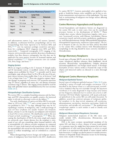
TABLE 28.1 Staging of Canine Mammary Tumors 15,125 for canine MGTs 129 ; however, particularly when applied to low-
grade or borderline lesions, some variability can be due to sub-
Lymph Node jective interpretation and experience of the pathologist. This may
VetBooks.ir Stage Tumor Size Status Metastasis lead to overreporting of malignant over benign tumors affecting
epidemiologic data.
Stage I
T1 <3 cm
Stage II T2 3–5 cm N 0 M 0
M 0
N 0
Canine Mammary Hyperplasia and Dysplasia
Stage III T3 >5 cm N 0 M 0
Several hyperplastic and dysplastic lesions are diagnosed in
Stage IV Any N 1 (positive) M 0 the canine MG and, in some cases, they can be considered
precursor lesions to the development of MGTs. 129 These
Stage V Any Any M 1 (metastasis)
include duct ectasia, lobular hyperplasia (regular, with secre-
tory [lactational] activity, with fibrosis [interlobular fibrous
connective tissue], and with atypia), epitheliosis, papillomato-
and subcutaneous tumors (e.g., mast cell tumors, lipomas). sis, and fibroadenomatous change (fibroepithelial hyperplasia,
In addition, correlations between cytopathology and ex vivo fibroepithelial hypertrophy, mammary hypertrophy). The lat-
histopathology have been reported to be between 68% and ter is frequent in the cat, but has not been clearly described
93%, 119,120 and the reported cytologic sensitivity and speci- in the canine MG; nodular lesions with fibroadenomatous
ficity for a malignant MGT diagnosis were 88% and 96%, morphology in the dog should be more correctly classified as
respectively. 121 Computed tomography (CT) imaging of the fibroadenomas.
thoracic cavity provides more sensitive detection of pulmonary
nodules than does thoracic radiography, but this may not be Benign Mammary Neoplasms
applicable for every patient because of increased expense and
limited availability. 121,122 Distant metastatic sites can include Several types of benign MGTs exist in the dog and include ade-
LNs, liver, lungs, and bone. 123 noma, intraductal papillary adenoma (duct papilloma), ductal
adenoma, fibroadenoma, myoepithelioma, complex adenoma
Staging System (adenomyoepithelioma), and benign mixed tumors. A histologic
MGTs are staged according to the T (tumor), N (lymph node), description of these various entities is beyond the scope of this
and M (metastasis) system. A modified version of the original chapter and interested readers are directed to a more thorough
staging system published by Owen 124 is currently used by most review. 129
oncologists: stage advances from I to II to III as the size of the pri-
mary tumor increases from smaller than 3 cm, to between 3 and Malignant Canine Mammary Neoplasms
5 cm, to larger than 5 cm. 125 These size categories capture impor-
tant changes in prognosis and outcome. LN metastasis represents Malignant Epithelial Tumors
stage IV disease, regardless of tumor size, and distant metastasis Several types of malignant epithelial tumors (Table 28.2) exist,
constitutes stage V disease. This staging system should be used for including a variety of simple and complex carcinomas. CIS is
dogs with epithelial tumors (noninflammatory) but not sarcomas described in the human breast as a well-demarcated, noninfil-
(Table 28.1). trative nodule(s) that has not extended through the basement
membrane; it is rarely diagnosed in dogs because early lesions
Histopathologic Classification Systems are less frequently detected than in women and criteria to dif-
The normal MG is a complex branching structure and the histo- ferentiate CIS from lobular hyperplasia with atypia and atypi-
logic and IHC characteristics are equally complex; the interested cal epitheliosis in the canine MG are still under discussion. In
reader is referred to a thorough review on the subject. 126 addition, several less common subtypes of malignant epithe-
Two early classifications of canine and feline MGTs were pub- lial neoplasm in the dog include squamous cell carcinomas,
lished in 1974 and 1999, 127,128 and a revised system for the dog adenosquamous carcinomas, mucinous carcinomas, lipid-rich
was published in 2011. 129 In dogs, although there may be his- carcinomas, and spindle cell carcinomas. 129 The predominant
topathologic evidence of malignancy, only a small percentage of morphologic pattern in each nodule is used to classify each
cases will have lymphatic and vascular invasion and metastatic nodule separately.
disease, whereas in cats the majority are malignant and metastasis
to local LNs is more common. The classification used in this text Malignant Mesenchymal Neoplasms: Sarcomas
is based on both morphology 129 and prognosis. 130,131 When dis- Malignant mesenchymal neoplasms of the MG most com-
cussing the classification of MGTs, the terms simple and complex monly include OSA, chondrosarcoma, fibrosarcoma, and
are commonly used. Simple denotes that the neoplasm is com- hemangiosarcoma. 129
posed of one cell type resembling either luminal epithelial cells OSA is the most common mesenchymal neoplasm of the
or myoepithelial cells, whereas complex neoplasms are composed canine MG and there is often a history of recent rapid growth
of two cell types, both luminal and myoepithelial cells, in which of a mammary mass that might have been present for some time.
the myoepithelial cells extend into the interstitium, show a typical A proliferation of cells varies from fusiform to stellate to ovoid,
spindle to stellate morphology, and are embedded in a myxoid and there is an association with islands of tumor osteoid and/or
matrix. 129 bone formation. Cartilage can also be present in variable amounts.
When distinguishing between benign and malignant tumors, Mitoses are frequently found. Metastasis occurs via the hematog-
criteria of malignancy have been listed in the 2011 classification enous route, mainly to the lungs. 