
Page 631 - Withrow and MacEwen's Small Animal Clinical Oncology, 6th Edition
P. 631
CHAPTER 28 Tumors of the Mammary Gland 609
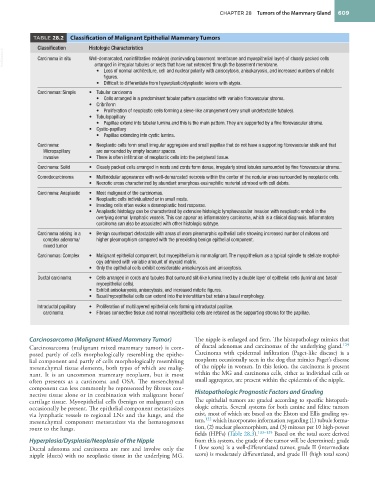
TABLE 28.2 Classification of Malignant Epithelial Mammary Tumors
Classification Histologic Characteristics
VetBooks.ir Carcinoma in situ Well-demarcated, noninfiltrative nodule(s) (noninvading basement membrane and myoepithelial layer) of closely packed cells
arranged in irregular tubules or nests that have not extended through the basement membrane.
• Loss of normal architecture, cell and nuclear polarity with anisocytosis, anisokaryosis, and increased numbers of mitotic
figures.
• Difficult to differentiate from hyperplastic/dysplastic lesions with atypia.
Carcinomas: Simple • Tubular carcinoma
• Cells arranged in a predominant tubular pattern associated with variable fibrovascular stroma.
• Cribriform
• Proliferation of neoplastic cells forming a sieve-like arrangement (very small undetectable tubules).
• Tubulopapillary
• Papillae extend into tubular lumina and this is the main pattern. They are supported by a fine fibrovascular stroma.
• Cystic-papillary
• Papillae extending into cystic lumina.
Carcinoma: • Neoplastic cells form small irregular aggregates and small papillae that do not have a supporting fibrovascular stalk and that
Micropapillary are surrounded by empty lacunar spaces.
invasive • There is often infiltration of neoplastic cells into the peripheral tissue.
Carcinoma: Solid • Closely packed cells arranged in nests and cords form dense, irregularly sized lobules surrounded by fine fibrovascular stroma.
Comedocarcinoma • Multinodular appearance with well-demarcated necrosis within the center of the nodular areas surrounded by neoplastic cells.
• Necrotic areas characterized by abundant amorphous eosinophilic material admixed with cell debris.
Carcinoma: Anaplastic • Most malignant of the carcinomas.
• Neoplastic cells individualized or in small nests.
• Invading cells often evoke a desmoplastic host response.
• Anaplastic histology can be characterized by extensive histologic lymphovascular invasion with neoplastic emboli in the
overlying dermal lymphatic vessels. This can appear as inflammatory carcinoma, which is a clinical diagnosis. Inflammatory
carcinoma can also be associated with other histologic subtype.
Carcinoma arising in a • Benign counterpart detectable with areas of more pleomorphic epithelial cells showing increased number of mitoses and
complex adenoma/ higher pleomorphism compared with the preexisting benign epithelial component.
mixed tumor
Carcinomas: Complex • Malignant epithelial component, but myoepithelium is nonmalignant. The myopithelium as a typical spindle to stellate morphol-
ogy admixed with variable amount of myxoid matrix.
• Only the epithelial cells exhibit considerable anisokaryosis and anisocytosis.
Ductal carcinoma • Cells arranged in cords and tubules that surround slit-like lumina lined by a double layer of epithelial cells (luminal and basal/
myoepithelial cells).
• Exhibit anisokaryosis, anisocytosis, and increased mitotic figures.
• Basal/myoepithelial cells can extend into the interstitium but retain a basal morphology.
Intraductal papillary • Proliferation of multilayered epithelial cells forming intraductal papillae.
carcinoma • Fibrous connective tissue and normal myoepithelial cells are retained as the supporting stroma for the papillae.
Carcinosarcoma (Malignant Mixed Mammary Tumor) The nipple is enlarged and firm. The histopathology mimics that
Carcinosarcoma (malignant mixed mammary tumor) is com- of ductal adenomas and carcinomas of the underlying gland. 129
posed partly of cells morphologically resembling the epithe- Carcinoma with epidermal infiltration (Paget-like disease) is a
lial component and partly of cells morphologically resembling neoplasm occasionally seen in the dog that mimics Paget’s disease
mesenchymal tissue elements, both types of which are malig- of the nipple in women. In this lesion, the carcinoma is present
nant. It is an uncommon mammary neoplasm, but it most within the MG and carcinoma cells, either as individual cells or
often presents as a carcinoma and OSA. The mesenchymal small aggregates, are present within the epidermis of the nipple.
component can less commonly be represented by fibrous con-
nective tissue alone or in combination with malignant bone/ Histopathologic Prognostic Factors and Grading
cartilage tissue. Myoepithelial cells (benign or malignant) can The epithelial tumors are graded according to specific histopath-
occasionally be present. The epithelial component metastasizes ologic criteria. Several systems for both canine and feline tumors
via lymphatic vessels to regional LNs and the lungs, and the exist, most of which are based on the Elston and Ellis grading sys-
mesenchymal component metastasizes via the hematogenous tem, 132 which incorporates information regarding (1) tubule forma-
route to the lungs. tion, (2) nuclear pleomorphism, and (3) mitoses per 10 high-power
fields (HPFs) (Table 28.3). 133–135 Based on the total score derived
Hyperplasia/Dysplasia/Neoplasia of the Nipple from this system, the grade of the tumor will be determined: grade
Ductal adenoma and carcinoma are rare and involve only the I (low score) is a well-differentiated tumor, grade II (intermediate
nipple (ducts) with no neoplastic tissue in the underlying MG. score) is moderately differentiated, and grade III (high total score)