
Page 45 - Veterinary Laser Therapy in Small Animal Practice
P. 45
Tissue healing 31
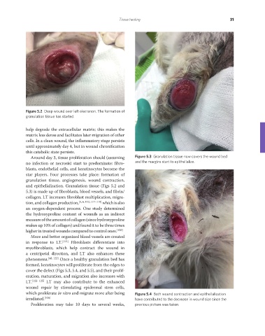
Figure 5.2 Deep wound over left olecranon. The formation of
granulation tissue has started.
help degrade the extracellular matrix; this makes the
matrix less dense and facilitates later migration of other
cells. In a clean wound, the inflammatory stage persists
until approximately day 4, but in wound chronification
this catabolic state persists.
Around day 3, tissue proliferation should (assuming Figure 5.3 Granulation tissue now covers the wound bed
no infection or necrosis) start to predominate: fibro- and the margins start to epithelialize.
blasts, endothelial cells, and keratinocytes become the
star players. Four processes take place: formation of
granulation tissue, angiogenesis, wound contraction,
and epithelialization. Granulation tissue (Figs 5.2 and
5.3) is made up of fibroblasts, blood vessels, and fibrin/
collagen. LT increases fi broblast multiplication, migra-
tion, and collagen production, [4, 6, 112, 117–119] which is also
an oxygen-dependent process. One study determined
the hydroxyproline content of wounds as an indirect
measure of the amount of collagen (since hydroxyproline
makes up 10% of collagen) and found it to be three times
higher in treated wounds compared to control ones. [120]
More and better organized blood vessels are created
in response to LT. [121] Fibroblasts differentiate into
myofibroblasts, which help contract the wound in
a centripetal direction, and LT also enhances these
phenomena. [48, 122] Once a healthy granulation bed has
formed, keratinocytes will proliferate from the edges to
cover the defect (Figs 5.3, 5.4, and 5.5), and their prolif-
eration, maturation, and migration also increases with
LT. [123–125] LT may also contribute to the enhanced
wound repair by stimulating epidermal stem cells,
which proliferate in vitro and migrate more after being Figure 5.4 Both wound contraction and epithelialization
irradiated. [126] have contributed to the decrease in wound size since the
Proliferation may take 10 days to several weeks, previous picture was taken.
REDONDO PRINT (4-COL BLEED).indd 31 08/08/2019 09:47