
Page 60 - Veterinary Laser Therapy in Small Animal Practice
P. 60
46 Veterinary Laser Therapy in Small Animal Practice
100% simulation data on a perfect slab of tissue with a per-
g=0.95 fectly cylindrical, homogeneous beam of photons inci-
g=0.6
10% g=0.3 dent perfectly perpendicular to the surface. And even in
that case you’d see some serious spreading of the beam
Probability 1% and decaying intensity over the first few centimeters of
depth. In fact, Figure 6.4 is based on some of this data.
[200]
But does that description of perfect geometry and
0.1%
homogeneous tissue sound like your Beagle’s hip? Not
even close. Also, do you plan to keep the hand-piece in
0.01% exactly the same spot for the entire treatment? If you’re
-180° -90° -0° 90° 180°
not too discouraged after this let-down of an answer to
Exit angle your main question and decide to keep reading, you’ll
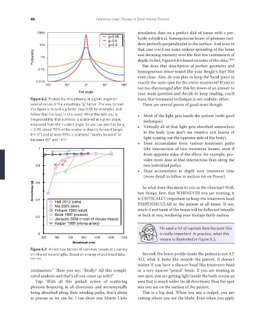
Figure 6.2 Probability of scattering at a given angle for learn that treatment technique is not realistic either.
several values of the anisotropy “g” factor. The way to read There are several pieces of good news though.
this figure is to pick a g factor (say 0.95 for example), and
follow that line (red, in this case). What this tells you is • Most of the light gets inside the patient (with good
the probability that a photon is scattered at a given angle, technique).
measured from the incident angle. So you can see that for g • Virtually all of that light gets absorbed somewhere
= 0.95, about 70% of the scatter is directly forward (angle
θ = 0°) and at least 99% is scattered “mostly forward” or in the body (you don’t see massive exit beams of
between 45° and −45°. light coming out the opposite side of the body).
• Dose accumulates from various treatment paths
1 (the intersection of two treatment beams, even if
from opposite sides of the elbow for example, pro-
vides more dose at that intersection than along the
0.8 two individual paths).
• Dose accumulates at depth over treatment time
(more detail to follow in section 6.6 on Power).
Anisotropy (g) two things: first, that WHENEVER you are treating, it
0.6
So what does this mean to you as the clinician? Well,
0.4
Hall 2012 (cells) is CRITICALLY important to keep the treatment head
PERPENDICULAR to the patient at all times. If not,
Ma 2005 (skin)
Firbank 1993 (skull) much if not most of the beam will be deflected laterally
Beek 1997 (muscle) or back at you, rendering your therapy fairly useless.
0.2
Jacques 2008 (mean of mouse tissue)
Keijzer 1989 (intima artery)
He used a lot of capitals here because this
is really important. In practice, what this
0
300 500 700 900 1100 1300 1500
means is illustrated in Figure 6.5.
Wavelength (nm)
Figure 6.3 Anisotropy factors of common tissues at a variety
of infrared wavelengths. Based on a range of published data. Second, the beam profile inside the patient is not AT
[194–199] ALL what it looks like outside the patient. It doesn’t
matter if you have a shower-head-like treatment head
centimeters.” Then you say, “Really? All this compli- or a very narrow “pencil” beam. If you are treating in
cated analysis and that’s all you come up with?” one spot, you are getting light inside the body across an
Yup. With all this pinball action of scattering area that is much wider (in all directions) than the spot
photons bouncing in all directions and incrementally size you see on the surface of the patient.
being absorbed along their winding paths, that’s about This is a big deal. When you use a scalpel, you are
as precise as we can be. I can show you Monte Carlo cutting where you see the blade. Even when you apply
REDONDO PRINT (4-COL BLEED).indd 46 08/08/2019 09:47