Page 163 - Veterinary Histology of Domestic Mammals and Birds, 5th Edition
P. 163
Blood and haemopoiesis (sanguis et haemocytopoesis)   145


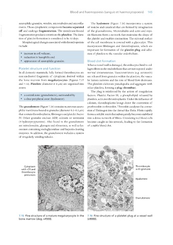
                  azurophilic granules, vesicles, microtubules and microfila-  The hyalomere (Figure 7.16) incorporates a system
       VetBooks.ir  ments. These cytoplasmic components become separated  of vesicles and canaliculi that are formed by invagination
                  off and undergo fragmentation. The membrane-bound  of the plasmalemma. Microtubules and actin and myo-
                                                                 sin filaments form a network that maintains the shape of
                  fragmentation products constitute the platelets. The dura-
                  tion of platelet formation is considered to be 12 days.  the platelet and enables contraction. The external surface
                     Morphological changes associated with thrombopoiesis  of the cell membrane is covered with a glycocalyx. This
                  include:                                       incorporates fibrinogen and thromboplastin, which are
                                                                 important for formation of the platelet plug and adhe-
                   ·  increase in cell volume,                   sion of platelets to the vascular endothelium.
                   ·  reduction in basophilia and
                   ·  appearance of azurophilic granules.        Blood clot formation
                                                                 When a vessel wall is damaged, thrombocytes bind to col-
                  Platelet structure and function                lagen fibres in the endothelium that are not exposed under
                  In all domestic mammals, fully formed thrombocytes are  normal circumstances. Vasoconstrictors (e.g. serotonin)
                  non-nucleated fragments of cytoplasm derived within  are released from granules within the platelets, the vascu-
                  the bone marrow from megakaryocytes (Figures 7.15  lar lumen narrows and the rate of blood flow decreases.
                  and 7.16). Platelets (diameter 2–4 μm) are organised into  The platelets elaborate pseudopodia and aggregate with
                  zones:                                         other platelets, forming a plug (thrombus).
                                                                    The plug is reinforced by the action of coagulation
                   ·  a central zone (granulomere), surrounded by   factors. Platelet Factor III, a phospholipid released by
                   ·  a clear peripheral zone (hyalomere).       platelets, activates thromboplastin. Under the influence of
                                                                 calcium, thromboplastin brings about the conversion of
                  The granulomere (Figure 7.16) contains numerous azuro-  prothrombin to thrombin. Thrombin catalyses the conver-
                  philic membrane-bound α-granules (diameter 0.2–0.3 μm)  sion of fibrinogen into the thread-like fibrin. Fibrin rapidly
                  that contain thromboplastin, fibrinogen and platelet Factor  forms a soluble mesh that subsequently becomes stabilised
                  IV.  Other  granules  enclose  ADP,  calcium  or  serotonin  into a dense network of fibres. Circulating red blood cells
                  (5-hydroxytryptamine). Also found in the granulomere  become caught in this network, leading to the formation
                  are mitochondria, glycogen and ribosomes, as well as lys-  of a stable blood clot.
                  osomes containing endoglycosidase and heparin-cleaving
                  enzymes. In addition, the granulomere includes a system
                  of irregularly winding tubules.


                  7.15  Fine structure of a mature megakaryocyte in the   7.16  Fine structure of a platelet plug at a vessel wall
                  bone marrow (dog; x4500).                      (x9000).


       Vet Histology.indb   145                                                                                  16/07/2019   14:58

