
Page 168 - Veterinary Histology of Domestic Mammals and Birds, 5th Edition
P. 168
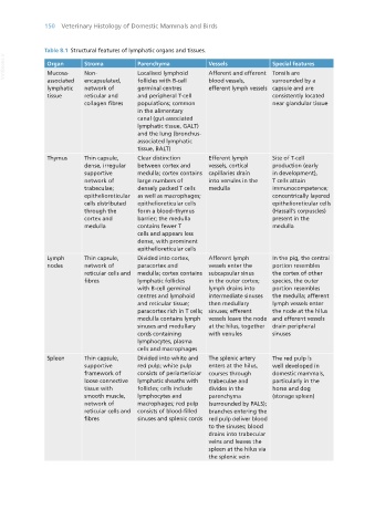
150  Veterinary Histology of Domestic Mammals and Birds


                   Table 8.1  Structural features of lymphatic organs and tissues.
       VetBooks.ir  Organ       Stroma           Parenchyma            Vessels              Special features

                                                 Localised lymphoid
                                Non-
                    Mucosa-
                                                                       Afferent and efferent  Tonsils are
                    associated   encapsulated,   follicles with B-cell   blood vessels,     surrounded by a
                    lymphatic   network of       germinal centres      efferent lymph vessels  capsule and are
                    tissue      reticular and    and peripheral T-cell                      consistently located
                                collagen fibres  populations; common                        near glandular tissue
                                                 in the alimentary
                                                 canal (gut-associated
                                                 lymphatic tissue, GALT)
                                                 and the lung (bronchus-
                                                 associated lymphatic
                                                 tissue, BALT)
                    Thymus      Thin capsule,    Clear distinction     Efferent lymph       Site of T-cell
                                dense, irregular   between cortex and   vessels, cortical   production (early
                                supportive       medulla; cortex contains  capillaries drain   in development),
                                network of       large numbers of      into venules in the   T cells attain
                                trabeculae;      densely packed T cells   medulla           immunocompetence;
                                epithelioreticular   as well as macrophages;                concentrically layered
                                cells distributed   epithelioreticular cells                epithelioreticular cells
                                through the      form a blood–thymus                        (Hassall’s corpuscles)
                                cortex and       barrier; the medulla                       present in the
                                medulla          contains fewer T                           medulla
                                                 cells and appears less
                                                 dense, with prominent
                                                 epithelioreticular cells
                    Lymph       Thin capsule,    Divided into cortex,   Afferent lymph      In the pig, the central
                    nodes       network of       paracortex and        vessels enter the    portion resembles
                                reticular cells and  medulla; cortex contains  subcapsular sinus   the cortex of other
                                fibres           lymphatic follicles   in the outer cortex;   species, the outer
                                                 with B-cell germinal   lymph drains into   portion resembles
                                                 centres and lymphoid   intermediate sinuses   the medulla; afferent
                                                 and reticular tissue;   then medullary     lymph vessels enter
                                                 paracortex rich in T cells;  sinuses; efferent   the node at the hilus
                                                 medulla contains lymph   vessels leave the node  and efferent vessels
                                                 sinuses and medullary   at the hilus, together   drain peripheral
                                                 cords containing      with venules         sinuses
                                                 lymphocytes, plasma
                                                 cells and macrophages
                    Spleen      Thin capsule,    Divided into white and   The splenic artery   The red pulp is
                                supportive       red pulp; white pulp   enters at the hilus,   well developed in
                                framework of     consists of periarteriolar  courses through   domestic mammals,
                                loose connective   lymphatic sheaths with   trabeculae and   particularly in the
                                tissue with      follicles; cells include   divides in the   horse and dog
                                smooth muscle,   lymphocytes and       parenchyma           (storage spleen)
                                network of       macrophages; red pulp   (surrounded by PALS);
                                reticular cells and  consists of blood-filled   branches entering the
                                fibres           sinuses and splenic cords  red pulp deliver blood
                                                                       to the sinuses; blood
                                                                       drains into trabecular
                                                                       veins and leaves the
                                                                       spleen at the hilus via
                                                                       the splenic vein


       Vet Histology.indb   150                                                                                  16/07/2019   14:59