Page 173 - Veterinary Histology of Domestic Mammals and Birds, 5th Edition
P. 173
Immune system and lymphatic organs (organa Iymphopoetica)  155


       VetBooks.ir


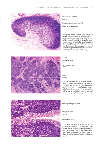
                                                                            8.8  Lymph node (sheep). The cortex is
                                                                            heavily populated with lymphocytes. It con-
                                                                            tains primary and secondary follicles. The
                                                                            lighter medulla consists largely of a loose
                                                                            arrangement of reticular and lymphoid
                                                                            tissue. Lymph passes from afferent vessels
                                                                            into the node via the subcapsular sinus and
                                                                            leaves through efferent vessels at the hilus.
                                                                            Haematoxylin and eosin stain (x15).


                                                                            8.9  Lymph node (pig). In this species,
                                                                            afferent lymph vessels enter the node at
                                                                            the hilus and leave via the subcapsular
                                                                            sinus. Therefore, lymph follicles aggre-
                                                                            gate in the inner zone of the node, with
                                                                            only a few present in the outer cortical
                                                                            zone. Haematoxylin and eosin stain (x20).


                                                                            8.10  Detailed view of a section of the
                                                                            cortex of the lymph node (dog). Reticular
                                                                            cell processes, macrophages and lympho-
                                                                            cytes are present within the subcapsular
                                                                            sinus, which continues into the interme-
                                                                            diate sinus. Haematoxylin and eosin stain
                                                                            (x720).


       Vet Histology.indb   155                                                                                  16/07/2019   14:59

