
Page 321 - Veterinary Histology of Domestic Mammals and Birds, 5th Edition
P. 321
Female reproductive system (organa genitalia feminina) 303
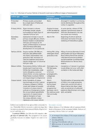
Table 14.1 Structure of ovarian follicles of domestic mammals at different stages of development.
VetBooks.ir Follicle type Oocyte Associated Special features
glandular activity
Primordial Primary oocyte surrounded None Arranged in rosettes in carnivores,
follicle by a single layer of flattened evenly distributed throughout the
follicular cells cortex in ungulates
Primary follicle Slight increase in volume Gradual increase In some species, release of FSH
of primary oocyte; oocyte in FSH secreted by induces a synchronous wave of
surrounded by single layer of adenohypophysis follicular development; the last
cuboidal follicular cells two waves are ovulatory
Secondary Substantial increase in size of Rise in FSH Beginning of antrum (antrum
follicle primary oocyte (80 μm), mitotic folliculare) formation through
division results in multilaminar widening of intercellular spaces
follicular cell stratum (5–10
layers); differentiation of stromal
cells into theca follicularis;
development of zona pellucida
Tertiary follicle Oocyte reaches 130–300 μm; Falling FSH, rising Filling of antrum (diameter 0.3 mm)
follicular cells differentiate oestrogens (from with protein-rich liquor follicularis;
into basal, intermediate and granulosa cells) theca interna forms from stromal
granulosa cells; formation of cells (steroid hormone synthesis)
cumulus oophorus and corona and spinocellular connective tissue
radiata (single layer of columnar differentiates into fibrous theca
follicular cells) externa
Mature follicle Preovulatory follicle; follicle and Oestrogens rise, Completion of meiosis I in primary
oocyte reach maximum size; FSH decreases, oocyte (does not occur until after
follicle lies immediately under secretion ovulation in the horse and dog)
tunica albuginea, avascular of LH and
stigma forms; cumulus oophorus prostaglandins,
floats freely in antrum; follicle collagenase
and stigma begin to degenerate activity
Corpus luteum Initial formation of corpus Rise in Transformation of granulosa cells
haemorrhagicum (after rupture progesterone of follicle into large granulosa
of stigma, collapse of follicle during luteal luteal cells and stromal cells of
lumen and release of oocyte); cell activity; theca interna into theca luteal
formation of corpus luteum progesterone cells; accumulation of yellowish
cyclicum or, if pregnancy occurs, decreases late in pigments (lipochromes; not in pigs
the corpus luteum graviditatis; the oestrous cycle; and ruminants)
regression of corpus luteum FSH eventually
leads to formation of scar- rises
like corpus albicans, corpus
nigrescens and corpus rubrum
follicle is surrounded by loose spinocellular connective tis- secondaRy follicles
sue. Primordial follicles represent a quiescent stage that Mitotic division of the follicular cells of a primary follicle
becomes reactivated to form primary follicles (Figures 14.3 leads to formation of the secondary follicle. Features of
to 14.5 and Table 14.1). secondary follicles (Table 14.1) include:
In primary follicles, the follicular cells comprise a sin-
gle layer of cuboidal cells. The cytoplasm of the oocyte · an increase in the volume of the oocyte (relative to
gradually increases in volume (Figures 14.1 and 14.3 to primary follicles),
14.5). · the zona pellucida,
· layering of the follicular cells and
· differentiation of stromal cells to form the theca
follicularis.
Vet Histology.indb 303 16/07/2019 15:05