
Page 323 - Veterinary Histology of Domestic Mammals and Birds, 5th Edition
P. 323
Female reproductive system (organa genitalia feminina) 305
VetBooks.ir
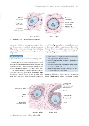
Flattened
follicular cell
follicular cell Cuboidal
Primary oocyte Primary oocyte
Basal lamina Basal lamina
Spinocellular
connective tissue
Spinocellular
connective tissue
Primordial follicle Primary follicle
14.5 Primordial and primary follicles (schematic).
In secondary follicles, the oocyte grows to 80 μm in diam- by follicular cell processes that are connected to the oocyte
eter. Concurrently, the cytoplasm undergoes structural by desmosomes and, later, by gap junctions. The primary
differentiation with an increase in protein-synthesising function of these intercellular connections is to provide
organelles (ER and ribosomes) and enlargement of the metabolic support to the oocyte (Figures 14.6 and 14.7).
Golgi complex. The zona pellucida:
Species variation · can be penetrated only by spermatozoa originating
Ruminants: The oocytes contain vacuolar lipid granules. from organisms of the same species,
· prevents the penetration of multiple spermatozoa
Cortical granules accumulate under the plasmalemma of (polyspermy),
the oocyte and the cell surface is irregularly studded with min- · counteracts premature implantation of the blasto-
ute cellular processes. The whole oocyte increases in size (this meres in the uterine tube,
continues until the early tertiary follicle stage) (Figure 14.6). · supports the structural integrity of the oocyte and
The zona pellucida is a fibrillar glycoprotein layer · regulates the uptake of nutrients.
(12–13 μm thick) that forms in the perivitelline space
between the surface of the oocyte and the adjacent fol- Secondary follicles are characterised by the prolifera-
licular cells (Figure 14.6). The zona pellucida is penetrated tion of follicular cells. Mitotic cell division gives rise
Follicular cells:
Basal cells
Intermediate cells
Granulosa cells
Follicular cell layers
Antrum
Oocyte
Corona radiata
Zona pellucida
Zona pellucida
Spinocellular Oocyte
connective tissue
Secondary follicle Tertiary follicle
14.6 Secondary and tertiary follicles (schematic).
Vet Histology.indb 305 16/07/2019 15:05