
Page 324 - Veterinary Histology of Domestic Mammals and Birds, 5th Edition
P. 324
306 Veterinary Histology of Domestic Mammals and Birds
VetBooks.ir
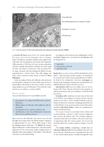
14.7 Fine structure of the zona pellucida and adjacent oocyte (sheep; x9000).
to several cell layers that encircle the oocyte (typically According to their location in the multilaminar wall of
five layers, up to ten in ruminants). Due to irregular the follicle (Figures 14.6, 14.8 and 14.9), the follicular cells
polar cell division, secondary follicles often appear oval. are designated as:
Individual cells are polygonal and contain few organelles.
Differentiation of follicular cells into an inner granulosa · basal cells,
cell layer, middle intermediate cell layer and outer basal · intermediate cells and
cell layer (see below) commences in the secondary folli- · granulosa cells.
cle stage. Stromal cells surrounding the follicle become
organised into a circular layer. The cells enlarge and Basal cells are in close contact with the basal lamina of the
form a thin connective tissue sheath termed the theca follicle. Their functions include transport of metabolites
follicularis. for nourishment of the oocyte and production of follicu-
In late secondary follicles, the follicular cells are loosely lar fluid. In the endoplasmic reticulum, oestrogens are
bound together. Expansion of intercellular spaces between formed from androgens (testosterone) synthesised by the
intermediate cells results in clefts that coalesce to form a epithelioid stromal cells of the theca interna.
larger cavity, the antrum folliculare. This marks the transi- Intermediate cells form the middle layer of the fol-
tion from a secondary to a tertiary follicle. licular wall. These cells are involved in the transport of
substances between cells and contribute to follicular fluid
teRtiaRy follicles production.
Characteristic features of tertiary follicles include: Granulosa cells constitute the inner layer of the wall
of the follicle, surrounding the antrum and the cumulus
· the presence of a large fluid-filled cavity (antrum oophorus (see below). These modified follicular cells syn-
folliculare), thesise protein-rich, hormone-containing antral fluid. In
· differentiation of follicular cells within the wall of tertiary follicles, many granulosa cells enclose PAS-positive
the follicle, granules (Call–Exner bodies) which are considered pre-
· development of the cumulus oophorus and cursors of follicular fluid.
· stratification of the theca follicularis to form the The oocyte is located eccentrically in the antrum and
theca interna and theca externa. is surrounded by a cluster of cells (cumulus oophorus)
that contacts the follicle wall. The cumulus oophorus
Spaces between the follicular cells can be detected with comprises basal cells, intermediate cells and an inner layer
the light microscope when the follicle reaches a diameter of (granulosa) cells, the corona radiata. The oocyte lies
of approximately 0.3 mm. These clefts merge to form centrally within the cumulus oophorus (Figures 14.8 and
the antrum (antrum folliculare). The fluid in the antrum 14.9).
(liquor follicularis) is rich in hyaluronic acid and protein. The corona radiata, comprising a simple layer of
Expansion of the antral cavity results in displacement of columnar follicular cells, lies immediately external to the
follicular cells to the periphery of the follicle. The follicular zona pellucida. Small processes extending from the cells
cells form a multi-layered lining. of the corona radiata project radially through the zona
Vet Histology.indb 306 16/07/2019 15:05