Page 329 - Veterinary Histology of Domestic Mammals and Birds, 5th Edition
P. 329
Female reproductive system (organa genitalia feminina)   311


                     From early in its formation, the corpus luteum is per-  Medulla (medulla ovarii, zona vasculosa)
       VetBooks.ir  vaded by a dense capillary network. The capillaries and  In most species, the medulla is the inner portion of the
                  hairpin arteries contribute to lobulation of the corpus  ovary enclosing blood vessels and nerves. Arteries enter
                  luteum. Connective tissue septa that subdivide the corpus  at the hilus and are arranged in coils within the medulla.
                  luteum arise from the theca externa and, to a lesser degree,  At the junction with the cortex, they form a plexus that
                  the theca interna. When the corpus luteum is actively  supplies the cortical tissue. These capillary networks are
                  secreting hormones, the capillary loops are in close contact  subject to cyclic structural changes regulated by specialised
                  with the luteal cells, facilitating the uptake of progesterone  arteries (‘barrier arteries’) and arteriovenous anastomoses.
                  into the blood circulation.                    Medullary lymph vessels and veins leave the ovary at the
                                                                 hilus. Predominantly autonomic nerve fibres innervate the
                  RegRession of the coRPus luteum                follicles and smooth muscle cells of the blood vessel walls.
                  Regression of the corpus luteum is initiated by prostaglan-
                  din (PGF ) secreted by the uterine mucosa. Hypertrophy   Species variation
                          2α
                  of blood vessel walls eventually results in blockage of the   Ruminants: Rete ovarii originating from the mesoneph-
                  lumen and degeneration of the capillary network. This is   ros are commonly present near the hilus. This is observed
                  accompanied by an increase in the amount of lipochrome   less frequently in other species. The rete ovarii consists of
                  pigments in the luteal cells and merging and enlargement   anastomosing cords and channels. These are composed of
                  of lipid vacuoles. The luteal cells undergo fatty degen-  epithelium-like cells and are surrounded by a basal lamina.
                  eration, with associated karyolysis, and are broken down   Horse: In the mare, the cortex lies internally, sur-
                  by macrophages. There is concurrent fibrosis (increase in   rounded by the medulla. Cortical tissue is only present
                  collagen fibres).                                on the surface of the ovary at the ovulation fossa.
                     In the ox, regression begins from about 15 days after
                  ovulation and becomes more rapid between days 18 and  Ovary of birds
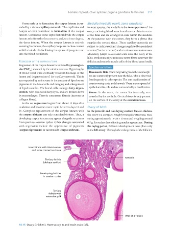
                  21. Complete replacement of the corpus luteum with  In the juvenile and non-laying mature female chicken,
                  the corpus albicans can take considerable time. Thus, a  the ovary is a compact, roughly triangular structure, mea-
                  developing corpus luteum may appear alongside structures  suring approximately 15–20 × 10 mm and weighing around
                  from previous ovarian cycles. Other changes associated  0.5 g. Its surface has a finely granular appearance. During
                  with  regression include the appearance of  pigments  the laying period, follicular development takes place only

                  (corpus nigrescens) or carotenoids (corpus rubrum).  in the left ovary. Through the enlargement of the follicles,


                  14.15  Ovary (chicken). Haematoxylin and eosin stain (x5).


       Vet Histology.indb   311                                                                                  16/07/2019   15:05

