
Page 334 - Veterinary Histology of Domestic Mammals and Birds, 5th Edition
P. 334
316 Veterinary Histology of Domestic Mammals and Birds
VetBooks.ir
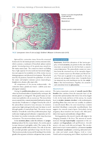
14.22 Low-power view of uterus (pig). Goldner’s Masson trichrome stain (x13).
Spinocellular connective tissue forms the structural Species variation
framework for the lamina propria (stroma endometrialis), Ruminants: Knob-like elevations of the lamina pro-
which contains a multitude of branched, tubular uterine pria, termed caruncles, are present in the cow. Similar
glands. Terminal portions of the glands may extend into structures are present in the ewe but have a concave
the tunica muscularis. The connective tissue scaffolding central depression. The spinocellular connective tissue
has a high capacity for structural and functional adapta- that forms the structural core of the caruncles (Figure
tion and supports the metabolic role of the uterine mucosa 14.21) contains numerous fibroblasts and blood ves-
during pregnancy and placental development. This is facili- sels. There are no glands in the caruncles. In the cow,
tated by the presence, beneath the epithelium, of cells of caruncles are arranged in four rows. Caruncles form
the innate and adaptive immune system (macrophages, the maternal mucosal attachment site for the cotyle-
lymphocytes, plasma cells, mast cells). dons of the fetal membranes. Together, the caruncles
All components of the stroma endometrialis – the and cotyledons form the placentome.
connective tissue, glands and vessels – exhibit cyclic mor-
phological variation. Myometrium
During the proliferation phase (pro-oestrus, oestrus), The tunica muscularis consists of smooth muscle fibre
when the hormonal milieu is dominated by oestrogen, the bundles that continue from the muscular layer of the
uterine glands are elongated. The gland lumina are nar- uterine tube and extend into the wall of the cervix (Figure
rowed and epithelial cells are numerous. The intercellular 14.20). In the body and horns of the uterus, the muscle
spaces expand; they contain interstitial fluid and are densely layer is circular (stratum circulare) with predominantly
vascularised. Production of collagen fibres by the cells of spiralling fibres that cross over one another. In addition
the spinocellular connective tissue increases. As ovulation to smooth muscle fibres, the outer muscle layer contains
approaches, high oestrogen levels cause the tissue to swell modified fibroblasts (contractile myofibroblasts) that
through uptake of intercellular fluid. The entire mucosa support the smooth muscle layers. Particularly during
thickens. Towards the end of the proliferation phase, high pregnancy, these transform into muscle cells. After partu-
molecular weight ground substance components are bro- rition they regress and synthesise collagen fibres.
ken down into smaller molecules and the tissue becomes During gestation, the smooth muscle cells adapt to the
oedematous. The microvasculature increases. changing demands of the fetus. The amount of muscle
In the secretory phase, under the influence of proges- increases through hypertrophy (expansion of the sar-
terone produced by the corpus luteum, the uterine glands coplasm) and hyperplasia (cell proliferation). Smooth
become shortened and tightly coiled. The lumina of the muscle cells may reach 800 μm in length. The circular-
glands expand and fill with secretion. The cells of the sim- spiral muscle layer is surrounded by a prominent vascular
ple cuboidal glandular epithelium bulge into the lumen. layer (stratum vasculosum) containing substantial arter-
Their secretory product is mucoid. Branching of the tubular ies, veins and lymph vessels (Figures 14.20 to 14.22).
glands is less apparent in bitches and queens than in mares. Vessels and accompanying nerve fibres (myelinated and
Vet Histology.indb 316 16/07/2019 15:05