Page 331 - Veterinary Histology of Domestic Mammals and Birds, 5th Edition
P. 331
Female reproductive system (organa genitalia feminina)   313


       VetBooks.ir


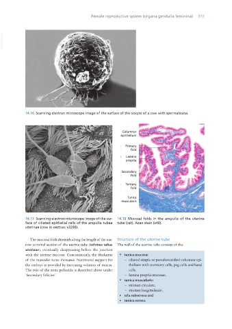
                  14.16  Scanning electron microscope image of the surface of the oocyte of a cow with spermatozoa.


                  14.17  Scanning electron microscope image of the sur-  14.18  Mucosal folds in the ampulla of the uterine
                  face of ciliated epithelial cells of the ampulla tubae   tube (cat). Azan stain (x40).
                  uterinae (cow in oestrus; x3200).


                     The mucosal folds diminish along the length of the nar-  Structure of the uterine tube
                  row terminal section of the uterine tube (isthmus tubae  The wall of the uterine tube consists of the:
                  uterinae), eventually disappearing before the junction
                  with the uterine mucosa. Concomitantly, the thickness   ·  tunica mucosa:
                  of the muscular tunic increases. Nutritional support for     − ciliated simple or pseudostratified columnar epi-
                  the embryo is provided by increasing volumes of mucus.   thelium with secretory cells, peg cells and basal
                  The role of the zona pellucida is described above under   cells,
                  ‘Secondary follicles’.                                − lamina propria mucosae,
                                                                   ·  tunica muscularis:
                                                                        − stratum circulare,
                                                                        − stratum longitudinale,
                                                                   •  tela subserosa and
                                                                   •  tunica serosa.


       Vet Histology.indb   313                                                                                  16/07/2019   15:05

