
Page 332 - Veterinary Histology of Domestic Mammals and Birds, 5th Edition
P. 332
314 Veterinary Histology of Domestic Mammals and Birds
Tunica mucosa The epithelium also contains peg cells. These are
VetBooks.ir The tunica mucosa (Figures 14.18 and 14.19) is an impor- thought to be cells that have become inactive after a secre-
tant functional component of the uterine tube. It is lined tory phase, or those that represent a transitional type.
with simple or pseudostratified columnar epithelium They are characterised by a typically pyknotic nucleus and
containing two main cell types: little cytoplasm. Basal cells lying close to the basal lamina
serve as reserve cells.
· ciliated cells and
· secretory cells. Tunica muscularis
The thickness of the tunica muscularis varies along the
The ciliated cells have motile cilia. Following ovulation, length of the uterine tube. In the infundibulum and
the secondary oocyte enters the ampulla and, in the early ampulla, the smooth muscle layers are thin. Circular and
phase of metoestrus, is sustained by the products of secre- isolated longitudinal or oblique muscle bundles are pres-
tory cells. Flow of fluid towards the uterus establishes ent without consistently discernible layering. The muscle
‘negative rheotactic’ movement of the spermatozoa, layer is markedly thicker in the isthmus and the fibre ori-
promoting their movement towards the ampulla. After entation is predominantly oblique. The inner component
fertilisation, the secretory capacity of the epithelium is is continuous with the circular muscle layer of the uterus.
maintained towards the uterus, ensuring sustained nour- Outer longitudinal muscle bundles radiate into the muscle
ishment of the developing embryo. of the tela subserosa of the uterus.
Rhythmic movement of the cilia facilitates the
movement of fluid and, thereby, the progression of the Tela subserosa and tunica serosa
zygote towards the uterus (during gestational dioestrus). Longitudinal muscle fibres extend through the tela sub-
Following implantation of the blastocyst in the uterine serosa of the isthmus tubae uterinae (lamina muscularis
mucosa, secretion by the cells of the uterine tube is sus- serosae). Blood vessels are present beneath the tela subse-
pended. If pregnancy does not occur, secretory activity rosa. A tunica serosa surrounds the uterine tube along its
resumes in subsequent pro-oestrus and oestrus. entire length.
Secretory cells release weakly acidic mucus and nutri-
ents (particularly proteins) that provide support for the Uterus
gametes. Secretion is by the merocrine mode. In addition, The uterus consists of a body (corpus uteri) connected to
secretory cells produce electrolytes, enzymes, albumins, the uterine tubes by the uterine horns (cornua uteri). The
glucose and amino acids required for final differentiation neck of the uterus, or cervix (cervix uteri), separates the
and maturation of the gametes. Both cell types undergo uterine body from the vagina (refer to Veterinary Anatomy
marked cyclic changes. of Domestic Mammals: Textbook and Colour Atlas). The uterus
and cervix are an integrated functional unit, and both are
subject to cyclic hormonal influences (refer to endocrinol-
ogy texts). The various functions of the uterus all serve
to support the reproductive process. Cyclic changes in
uterine structure (proliferation, secretion, involution) are
mediated by endocrine regulatory mechanisms and form
part of the overall reproductive cycle (see below, under
‘Oestrous cycle’; refer also to endocrinology texts).
The surface epithelium and smooth muscle of the
uterus facilitate transport of spermatozoa towards the
uterine tubes. This is also influenced by cyclic events.
Uterine secretions contribute to capacitation (final mat-
uration) of spermatozoa which enables spermatozoa
to penetrate the cells of the corona radiata and the zona
pellucida, allowing fertilisation to occur.
The uterus supports the developing embryo and serves
to expel the placenta at the end of pregnancy. Cyclic modi-
fications of the uterine mucosa are necessary to provide
optimal conditions for implantation and formation of the
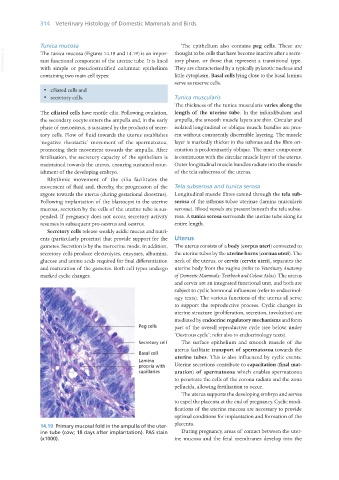
14.19 Primary mucosal fold in the ampulla of the uter- placenta.
ine tube (cow; 18 days after implantation). PAS stain During pregnancy, areas of contact between the uter-
(x1000). ine mucosa and the fetal membranes develop into the
Vet Histology.indb 314 16/07/2019 15:05