
Page 316 - Veterinary Histology of Domestic Mammals and Birds, 5th Edition
P. 316
298 Veterinary Histology of Domestic Mammals and Birds
VetBooks.ir
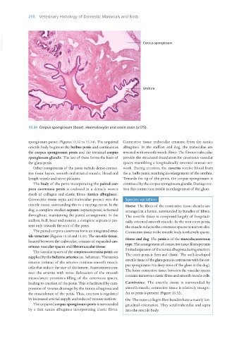
13.34 Corpus spongiosum (boar). Haematoxylin and eosin stain (x175).
spongiosum penis) (Figures 13.32 to 13.34). The unpaired Connective tissue trabeculae emanate from the tunica
erectile body begins as the bulbus penis and continues as albuginea. In the stallion and dog, the trabeculae are
the corpus spongiosum penis and the terminal corpus invested with smooth muscle fibres. The fibrous trabeculae
spongiosum glandis. The last of these forms the basis of provide the structural foundation for cavernous vascular
the glans penis. spaces resembling a longitudinally oriented venous net-
Other components of the penis include dense connec- work. During erection, the caverns receive blood from
tive tissue layers, smooth and striated muscle, blood and the a. bulbi penis, resulting in enlargement of the urethra.
lymph vessels and nerve plexuses. Towards the tip of the penis, the corpus spongiosum is
The body of the penis incorporating the paired cor- continued by the corpus spongiosum glandis. During erec-
pora cavernosa penis is enclosed in a densely woven tion this connection results in enlargement of the glans.
mesh of collagen and elastic fibres (tunica albuginea).
Connective tissue septa and trabeculae project into the Species variation
erectile tissue, surrounding this to a varying extent. In the Horse: The fibres of the connective tissue sheaths are
dog, a complete median septum (septum penis) is formed arranged in a lattice, surrounded by bundles of fibres.
throughout, maintaining the paired arrangement. In the The erectile tissue is composed largely of longitudi-
stallion, bull, boar and tomcat, a complete septum is pre- nally oriented smooth muscle. In the non-erect penis,
sent only towards the root of the penis. the muscle reduces the cavernous spaces to narrow slits.
The paired corpora cavernosa form an integrated erec- Connective tissue in the erectile body is relatively sparse.
tile structure (Figures 13.32 and 13.33). The erectile tissue,
located between the trabeculae, consists of expanded cav- Horse and dog: The penis is of the musculocavernous
ernous vascular spaces and fibromuscular tissue. type. The arrangement of connective tissue fibres permits
The vascular spaces of the corpora cavernosa penis are limited expansion of the tunica albuginea during erection.
supplied by the helicine arteries (aa. helicinae). The tunica The erect penis is firm and elastic. The well-developed
interna (intima) of the arteries contains smooth muscle erectile tissue of the glans penis is continuous with the cor-
cells that reduce the size of the lumen. Anastomoses con- pus spongiosum (via deep veins of the glans in the dog).
nect the arteries with veins. Relaxation of the smooth The loose connective tissue between the vascular spaces
musculature promotes filling of the cavernous spaces, contains numerous elastic fibres and smooth muscle cells.
leading to erection of the penis. This is facilitated by com- Carnivores: The erectile tissue is surrounded by
pression of venous drainage by the tunica albuginea and smooth muscle; connective tissue is relatively meagre.
the musculature of the penis. Thus, erection is regulated An os penis is present (Figure 13.32).
by increased arterial supply and reduced venous outflow. Ox: The outer collagen fibre bundles have a mainly lon-
The unpaired corpus spongiosum penis is surrounded gitudinal orientation. They send trabeculae and septa
by a thin tunica albuginea incorporating elastic fibres. into the erectile body.
Vet Histology.indb 298 16/07/2019 15:05