
Page 69 - Veterinary Histology of Domestic Mammals and Birds, 5th Edition
P. 69
Epithelial tissue (textus epithelialis) 51
prostaglandins) (Figure 2.25). This form of secretion is elements of exocrine glands is provided in Table 2.3. Based
VetBooks.ir referred to as paracrine. It is employed, for example, by on their relationship to the surface epithelium, exocrine
glands are classified as:
enteroendocrine cells of the gastro-intestinal tract.
In the autocrine mode of secretion, the hormone acts
upon the cell by which it was secreted (e.g. Leydig cells in • intra-epithelial glands (glandulae intraepitheliales)
the testes) (see also Chapter 9, ‘Endocrine system’). or
• extra-epithelial glands (glandulae exoepitheliales).
Exocrine glands (glandulae exocrinae)
Exocrine glands are glands in which the secretory product INTRA-EPITHELIAL GLANDS (GLANDULAE
is transported to the surface of the epithelium (Figures 2.31 INTRAEPITHELIALES)
to 2.44). Consisting of mucin, enzymes or other materials, Intra-epithelial glands may consist of a single cell or a
the secretion reaches its destination in one of two ways: by group of cells.
passing directly to the internal or external body surface, or Goblet cells are typical single-cell intra-epithelial exo-
by travelling through a single duct or complex system of crine glands (Figures 2.26 to 2.30). They are club-shaped
ducts. The composition and concentration of the secretory cells with a narrow base. The secretory product, mucino-
product may undergo modification during its passage to gen, is synthesised by the endoplasmic reticulum and Golgi
the surface. Exocrine glands include the mammary, sali- apparatus, and accumulates in the apical region of the
vary and bronchial glands. A summary of the structural cell in the form of membrane-bound secretory granules.
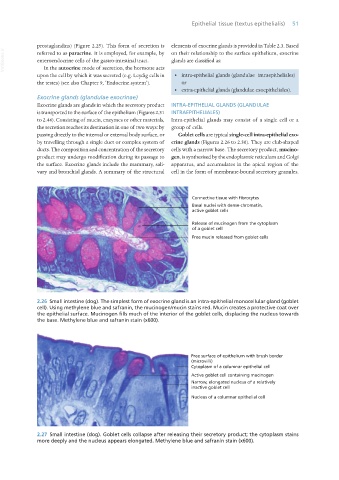
2.26 Small intestine (dog). The simplest form of exocrine gland is an intra-epithelial monocellular gland (goblet
cell). Using methylene blue and safranin, the mucinogen/mucin stains red. Mucin creates a protective coat over
the epithelial surface. Mucinogen fills much of the interior of the goblet cells, displacing the nucleus towards
the base. Methylene blue and safranin stain (x600).
2.27 Small intestine (dog). Goblet cells collapse after releasing their secretory product; the cytoplasm stains
more deeply and the nucleus appears elongated. Methylene blue and safranin stain (x600).
Vet Histology.indb 51 16/07/2019 14:54