
Page 72 - Veterinary Histology of Domestic Mammals and Birds, 5th Edition
P. 72
54 Veterinary Histology of Domestic Mammals and Birds
VetBooks.ir
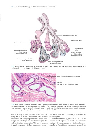
2.33 Serous, mucous and mixed secretory units of a compound tubulo-acinar gland with myoepithelial cells
(schematic). See also Chapter 10, ‘Digestive system’.
2.34 Sweat gland, skin (calf). Sweat glands are typically simple coiled tubular glands. In this histological section,
several cross-sections of the same gland are evident. The gland is lined by a single layer of cuboidal epithelium
that produces a watery secretion (sweat). Sweat glands are classified as extra-epithelial, exocrine, tubular
merocrine or apocrine glands. Haematoxylin and eosin stain (x250).
content of the granules is released at the cell surface by mechanism and occurs in the exocrine pancreas and in the
exocytosis. In this process, the membrane of the secretory endocrine glands.
vesicle fuses with the apical plasmalemma and the secre- In apocrine secretion (Figures 2.35, 2.36 to 2.38), the
tory product is discharged into the extracellular space. The secretory granule is pinched off from the free cell surface
cell structure thus remains intact. This type of secretion together with a small amount of cytoplasm. Secretory
is usually continuous. It is the most common secretory granules are formed in the supranuclear region of the
Vet Histology.indb 54 16/07/2019 14:54