
Page 71 - Veterinary Histology of Domestic Mammals and Birds, 5th Edition
P. 71
Epithelial tissue (textus epithelialis) 53
shaPe of the secRetoRy unit Compound glands are usually divided into lobules
VetBooks.ir 2.31 to 2.33) may be described as: specific structure. The secretory product enters a complex
Based on the shape of the lumen, secretory units (Figures containing groups of secretory units that exhibit an organ-
system of strongly branching ducts, before emptying into
• tubular (resembling a hose or pipe), a single excretory duct. This type of gland is exemplified
• acinar (spherical, berry-shaped) or by the large salivary glands (Figure 2.33).
• alveolar (vesicular) or
• mixed types, such as tubulo-alveolar or tubulo- mode of secRetion
acinar (often associated with compound glands). The various modes in which the secretory product is
released from the epithelial cells include:
The walls of the secretory units are comprised of secre- • merocrine secretion (also referred to as eccrine),
tory cells. Generally, the secretory end pieces consist of • apocrine secretion and
a single layer of glandular epithelium. Sebaceous glands, • holocrine secretion.
in which multiple cell layers are present, are an exception
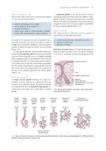
(Figure 2.39). Merocrine secretion (Figure 2.37) involves the release of
In some glands, the base of the secretory units is sur- substances that have been stored in the apical cytoplasm
rounded by myoepithelial cells that function as contractile in the form of membrane-bound secretory granules. The
elements. Embryologically, these are modified epithelial
cells containing bundles of myofilaments. These cells may
be arranged in parallel, or as a branching network, adjacent
to the basal surface of the cells. Contraction of myoepithe-
lial cells assists in expelling the secretory product into the
duct. These cells are found particularly in sweat glands, the
alveolae of the mammary glands and the salivary glands
(Figure 2.33).
stRuctuRe of the ducts
In simple exocrine glands consisting of a single mul-
ticellular secretory unit and an unbranched duct, the
secretory unit is typically tubular. Examples include the
sweat glands of the skin. In branched simple glands, sev-
eral secretory units empty into a single duct (e.g. gastric 2.32 Acinar and tubular secretory units with associ-
glands). ated duct (schematic).
2.31 Types of exocrine tubular and alveolar glands (schematic), illustrating varying degrees of differentiation
(simple, simple branched, compound).
Vet Histology.indb 53 16/07/2019 14:54