Page 227 - Manual of Equine Field Surgery
P. 227
Urethral Extension (Urethroplasty) 223
-,.~
r----- ·----·
-
..:.;,,._
I
I
A
B
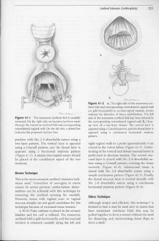
Figure 41-2 A, The right side of the transverse ure-
thral fold and corresponding ventrolateral vaginal wall
are split horizontally i11 an interrupted manner. Arrows
indicate the direction of tissue mobilization. The left
Figure 41-1 The transverse urethral fold is caudally side of the transverse urethral fold has been sutured to
retracted. On the right side, an incision has been made the corresponding ventrolateral vaginal wall. B, Close-
through the transverse urethral fold and corresponding t1p view of a two-layer closure. The ventral layer is
ventrolateral vaginal wall. On the left side, a dotted line apposed using a ConneU pattern, and the dorsal layer is
indicates the proposed incision line. apposed using a continuous horizontal mattress
pattern.
position with No. 2-0 absorbable suture using a
two-layer pattern. The ventral layer is apposed right vaginal walls to a point approximately 3 cm
using a Connell pattern, and the dorsal layer is cranial to the vulvar labiae (Figure 41-3). Under-
apposed using a horizontal mattress pattern mining of the ventral and dorsal mucosal layers is
(Figure 41-2). A simple interrupted suture should performed to decrease tension. The ventral mu-
be placed at the caudalmost aspect of the two cosal layer is closed with No. 2-0 absorbable su-
•
111ClSIOnS. ture using a Connell pattern, everting the tissue
ventrally (Figure 41-4). Submucosal tissue is
closed with No. 2-0 absorbable suture using a
Brown Technique
simple continuous pattern (Figure 41-5). Finally,
This is the most common urethral extension tech- the dorsal mucosal layer is everted dorsally with
3
nique used. Correction of urovagina in mares No. 2-0 absorbable suture using a continuous
caused by severe perinea! conformation abnor- horizontal mattress pattern (Figure 41-6).
malities can be achieved with this technique by
extending the urethral opening far caudally. Shires Technique
However, rnares with vaginal scars or vaginal
mucosa atrophy are not good candidates for this Although simple and efficient, this technique" is
technique because of increased tissue tension.3 limited in that it may be used only in mares that
A 30- Fr Foley catheter is placed in the urinary have redundant vestibular folds that may be
bladder and the cuff is inflated. The transverse pulled together to form a tunnel without the need
urethral fold is split horizontally, and the mucosal for dissecting and undermining tissue flaps to
1
incision is extended caudally along the left and form a shelf.

