Page 229 - Manual of Equine Field Surgery
        P. 229
     '                                                                                                                                                                       Urethral Extension  (Urethroplasty)                                               225
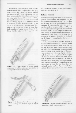
                                 A 30-Fr  Foley catheter is  placed  in the urinary                                                                   No. 2-0 absorbable  suture  using  a simple  contin-
                          bladder  and the  cuff  is  inflated.  Before any inci-                                                                     uous pattern  (Figure  41-9).
                          sion  is  made,  two  lines  of  the  ventral  vaginal
                           mucosa are  dorsally everted and sutured  over the                                                                         McKinnon Technique
                           Foley catheter with No. 0 absorbable suture using
                           a11  interrupted  horizontal  mattress  pattern,                                                                           Correction  of urovagina  in mares  caused by severe
                           leaving  adequate  mucosa  to  allow  excision  and                                                                        perineal  conformation  abnormalities                                                        can  be
                           further suturing  (Figure  41-7).  Suture placement                                                                        accomplished  with this technique=  by providing
                           is  continued  caudally  to  approximately  2  cm                                                                          a wide, long,  and  strong  urethral  extension.  This
                           cranial to the vulvar labiae.  The two lines of dor-                                                                       technique  is  recommended  when  the  urethra
                           sally  everted  mucosa  are  excised  to  create  four                                                                     opening  needs  to  be  extended  far  caudally  and
                           fresh-cut  edges  of vaginal  mucosa  (Figure 41-8).                                                                       increased  tissue tension  is present.  Minimal  tissue
                           These  debrided  edges  are  then  apposed  with                                                                           tension  is exerted  on the completed  tunnel.  I11i-
                                                                                                                                                      tially, a steep  learning  curve for this  technique  is
                                                                                                                                                      encountered,  but it can  be  easily performed with
                                                                                                                                                      experience.  In  addition,  disruption  of  the  blood
                                                                                                                                                      supply  should  be  avoided  during  the  tissue  flap
                                                                                                                                                      dissection.
                                                                                                                                                             A 30-Fr  Foley catheter  is placed  in the  urinary
                                                                                                                                                      bladder  and the cuff is inflated, The caudal border
                                                                                                            '                                          of  the  transverse  urethral  fold  is  grasped  on
                                                                                                                                                      midline  with  Allis  tissue  forceps  and  retracted
                                                                                                                                                       caudally. A horizontal  mucosal  incision  is made 2
                                                                                                                                                       to  4 cm  cranial  to  the  caudal  edge  of the  trans-
                                                                                                                                                       verse  urethral  fold  extending  slightly  dorsocau-
                                                                                                                                                       dally along the left and  right vaginal walls (Figure
                                                                                                                                                       41-10).           This  incision  should  end  at  the  vulvar
                                                                                                                                                       labia half to two thirds  of the distance  between  the
                                                                                                                                                       vaginal floor  and vaginal roof. The transverse ure-
                                                                                                                                                       thral  fold  and  vaginal  wall  mucosal  tissues  are
                                                                                                                                                       undermined  so  that  the  free  tissue  flaps  are
                            Figure  41-7                      Dorsal  eversion  of  ventral  vaginal                                                   reflected  caudally  and  axially,  respectively.  Dis-
                            mucosa over 30-Fr  Foley catheter  and sutured using an                                                                    section  of transverse  urethral  fold  tissue  should
                            interrupted  horizontal  mattress  pattern.
                             Figure  41-8                     Excision  of  dorsally  everted  vaginal
                             mucosa,  creating  four  fresh-cut  edges  of  vaginal                                                                     Figure 41-9  Apposition  of freshly debrided  mucosa!
                             mucosa.                                                                                                                    edges using a simple  continuous  pattern.


