
Page 695 - Clinical Small Animal Internal Medicine
P. 695
61 Imaging in Hepatobiliary Disease 663
the gallbladder wall may be recognized as fine curvilin- the ultrasound probe is placed in the midline just caudal
VetBooks.ir ear mineralization and choleliths are typically seen as to the xiphisternum and the ultrasound beam is “swept”
through the liver in both sagittal and transverse planes.
multiple small mineralized opacities superimposed on
the right cranioventral liver. Branching mineralization is
gins (typically deep‐chested patients and those with a
occasionally recognized as an apparently incidental find- In patients where the liver lies well within the costal mar-
ing in older terrier dogs. The identification of gas opacity small liver), it can be challenging to get a good image
within the liver is even less common but may be seen as from a subcostal window, and it is often easier to exam-
streaks or patches of gas opacity (e.g., in patients with ine each side of the liver by obtaining dorsal and trans-
emphysematous cholecystitis or abscessation) or as verse plane images through the adjacent intercostal
branching lucencies due to gas migrating throughout the spaces. The gallbladder, common bile duct, and caudal
biliary tree (e.g., due to emphysematous cholecystitis) or, vena cava all lie to the right of midline; for this reason,
very rarely, throughout the portal venous system. evaluation of the biliary tract and screening for possible
portosystemic shunts are often performed with the
Changes in Margination transducer positioned on the right side, either just caudal
Clearly defined ventral and caudoventral hepatic mar- to the costal arch or within the caudal intercostal spaces.
gins rely on the presence of abdominal fat highlighting
the soft tissue opacity of the liver; loss of this clear Recognizing Artifacts
margination may be seen in patients with ascites and in
very young or emaciated patients. Several artifacts are commonly encountered during
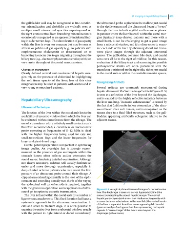
hepatic ultrasound. The “mirror‐image” artifact (Figure 61.5)
is seen as a reflection of the liver beyond the diaphragm,
Hepatobiliary Ultrasonography and is caused by the highly reflective interface between
the liver and lung. “Acoustic enhancement” is caused by
the fact that fluid results in less attenuation of the ultra-
Ultrasound Technique
sound beam than soft tissues, and is recognized as the
The location of the liver within the costal arch limits the tissues deep to a fluid‐filled structure, such as the gall-
availability of acoustic windows from which the liver can bladder, appearing artificially echogenic relative to the
be evaluated without interference from the ribcage. The adjacent tissues.
use of a transducer with a relatively small contact area is
therefore recommended; a curvilinear or microconvex
probe operating at frequencies of 5–12 MHz is ideal,
with the higher frequencies being used for cats and
small‐to‐medium dogs and the lower frequencies for
large‐ and giant‐breed dogs.
Careful patient preparation is important in optimizing
image quality. An overnight fast is strongly recom-
mended, as the presence of gas and ingesta within the
stomach lumen often reflects and/or attenuates the
sound waves, hindering detailed examination. Although
not always necessary, sedation will usually facilitate an
easier and more thorough examination, especially in
deep‐chested or tense patients who may resent the firm
pressure of an ultrasound probe around their ribcage. A
clipped area extending cranially to the level of the xiphi-
sternum and extending dorsally two‐thirds of the way up
the abdominal wall on either side is required, together
with the generous application and reapplication of ultra- Figure 61.5 A sagittal plane ultrasound image of a normal canine
sound gel to optimize acoustic transmission. liver. The diaphragm is seen as a curved hyperechoic line (blue
The liver is fixed within the costal arch by a number of arrows) demarcating the cranial hepatic margin. The normal
ligamentous attachments. This fixed location facilitates a hepatic parenchyma (pink arrow) is of medium echogenicity with
systematic approach to the ultrasound examination. In a coarse but even echotexture. In the near field, the ventral border
cats and small‐to‐medium dogs, it is often possible to of the liver is separated from the coarser appearing falciform fat
(green arrow) by a fine hyperechoic line representing the hepatic
examine the entire liver from a subcostal window, usually capsule. A “mirror‐image” of the liver is seen beyond the
with the patient in right lateral or dorsal recumbency: diaphragm (yellow arrow).