
Page 699 - Clinical Small Animal Internal Medicine
P. 699
61 Imaging in Hepatobiliary Disease 667
may be acoustic enhancement deep to fluid content, with of serosal detail will be seen on abdominal radiographs
VetBooks.ir occasional gas bubbles recognized as brightly echogenic due to the presence of free fluid. Depending on the vol-
ume of fluid, it may also be possible to appreciate altera-
foci with distal reverberation artifact. Mineralization of
the abscess capsule may result in echogenic foci with dis-
tal acoustic shadowing. Most hepatic abscesses will not tion of the normal hepatic shape.
be radiographically apparent; however, lesions bulging Neoplastic Disease
from the liver surface and lesions with mineralization of
the capsule and/or gas within the abscess cavity may be The WSAVA classification of neoplastic disorders
identified. Concurrent hepatic lymphadenopathy, abnor- includes primary neoplasia, usually arising from the
mally hyperechoic local mesentery, and free abdominal hepatocytes (hepatocellular adenoma/carcinoma) and/or
fluid may be seen in some patients; radiographically, the biliary system (e.g. cholangiocellular adenoma/carci-
these features would be recognized as a progressive loss noma), primary vascular neoplasia (most commonly
of serosal detail. hemanigosarcoma), diffusely infiltrative hematopoietic
neoplasia (e.g., lymphoma, malignant histiocytosis, and
Cystic Lesions malignant mastocytosis) and metastatic neoplasia.
Hepatic cysts are recognized as well‐circumscribed, Although benign, nodular hyperplasia, commonly seen in
single or multiple, uni‐ or multilocular, thin‐walled older dogs but less frequently identified in cats, is also
structures, with anechoic contents and distal acoustic included in the classification of neoplastic disease.
enhancement. Congenital cystic diseases of the dog and
cat are occasionally seen and may be associated with Primary Focal Neoplasia
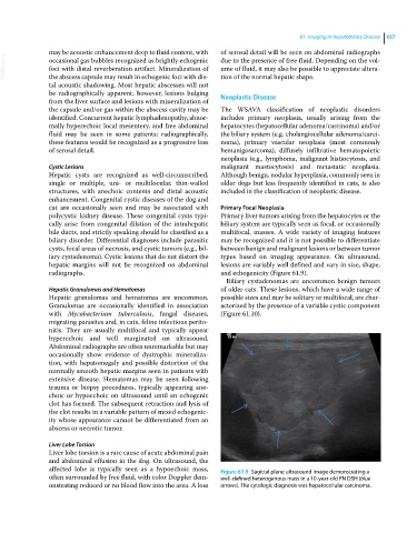
polycystic kidney disease. These congenital cysts typi- Primary liver tumors arising from the hepatocytes or the
cally arise from congenital dilation of the intrahepatic biliary system are typically seen as focal, or occasionally
bile ducts, and strictly speaking should be classified as a multifocal, masses. A wide variety of imaging features
biliary disorder. Differential diagnoses include parasitic may be recognized and it is not possible to differentiate
cysts, focal areas of necrosis, and cystic tumors (e.g., bil- between benign and malignant lesions or between tumor
iary cystadenoma). Cystic lesions that do not distort the types based on imaging appearance. On ultrasound,
hepatic margins will not be recognized on abdominal lesions are variably well defined and vary in size, shape,
radiographs. and echogenicity (Figure 61.9).
Biliary cystadenomas are uncommon benign tumors
Hepatic Granulomas and Hematomas of older cats. These lesions, which have a wide range of
Hepatic granulomas and hematomas are uncommon. possible sizes and may be solitary or multifocal, are char-
Granulomas are occasionally identified in association acterized by the presence of a variable cystic component
with Mycobacterium tuberculosis, fungal diseases, (Figure 61.10).
migrating parasites and, in cats, feline infectious perito-
nitis. They are usually multifocal and typically appear
hyperechoic and well marginated on ultrasound.
Abdominal radiographs are often unremarkable but may
occasionally show evidence of dystrophic mineraliza-
tion, with hepatomegaly and possible distortion of the
normally smooth hepatic margins seen in patients with
extensive disease. Hematomas may be seen following
trauma or biopsy procedures, typically appearing ane-
choic or hypoechoic on ultrasound until an echogenic
clot has formed. The subsequent retraction and lysis of
the clot results in a variable pattern of mixed echogenic-
ity whose appearance cannot be differentiated from an
abscess or necrotic tumor.
Liver Lobe Torsion
Liver lobe torsion is a rare cause of acute abdominal pain
and abdominal effusion in the dog. On ultrasound, the
affected lobe is typically seen as a hypoechoic mass, Figure 61.9 Sagittal plane ultrasound image demonstrating a
often surrounded by free fluid, with color Doppler dem- well‐defined heterogenous mass in a 10‐year‐old FN DSH (blue
onstrating reduced or no blood flow into the area. A loss arrows). The cytologic diagnosis was hepatocellular carcinoma.