
Page 701 - Clinical Small Animal Internal Medicine
P. 701
61 Imaging in Hepatobiliary Disease 669
Extrahepatic Cholestasis lymphocytic cholangitis are both commonly diagnosed
VetBooks.ir include inflammation or neoplasia of the common bile in cats, but less frequently reported in dogs. On ultra-
Causes of extrahepatic biliary obstruction (EHBO)
sound, cholangitis is most commonly recognized as
duct, pancreas or duodenum and obstruction of the duct
lights the echogenic walls of the portal vasculature
lumen by choleliths (choledocholithiasis). Dilation of the diffusely hypoechoic hepatic parenchyma, which high-
common bile duct (>5 mm in cats and >4 mm in dogs) is (Figure 61.13).
the most consistent ultrasound finding in acute EHBO These changes are nonspecific, and it is not possible to
(Figure 61.12). make a definitive diagnosis or to distinguish between
Dilation of the extrahepatic and intrahepatic bile ducts neutrophilic and lymphocytic disease without tissue
may be seen in patients with obstruction of at least 5–7 sampling. Cholangitis cannot be diagnosed from abdom-
days duration; these dilated ducts are recognized as the inal radiographs. Concurrent cholecystitis (see later) is
presence of multiple slightly tortuous anechoic tubes identified in a significant number of cats and occasion-
with hyperechoic walls, which do not show any evidence ally dogs with cholangitis.
of flow on Doppler. Dilation of the gallbladder is an
inconsistent finding and the absence of a distended gall- Gallbladder Mucocele
bladder does not exclude the possibility of EHBO. Gallbladder mucoceles occur in dogs and are caused by
Detailed evaluation of the common bile duct is required an abnormal accumulation of inspissated mucus and bile
to identify possible causes of obstruction; obstructive within the gallbladder lumen, leading to progressive
choleliths are most commonly located in the distal com- luminal distension and potentially to necrosis and ulti-
mon bile duct, highlighted by the dilated bile duct proxi- mately rupture of the gallbladder wall. Typical mucoceles
mal to the obstruction, with an empty bile duct seen have a distinctive ultrasound appearance, described as
distally. In patients with inflammatory or neoplastic dis- “kiwi‐like” or “stellate,” with immobile hyperechoic stria-
ease, an extraluminal mass may be identified causing tions seen radiating from a central point (Figure 61.14).
obstruction; however, it is not possible to determine the Variations in the appearance include a “mosaic‐like”
nature of the mass (inflammatory or neoplastic) from the pattern of mixed echogenicity; unlike gallbladder sludge,
ultrasound appearance. Although mineralized choleliths which is a common and often incidental finding, the con-
may be identified on abdominal radiographs, EHBO will tents of a gallbladder mucocele are immobile and do not
not be seen. move with gravity when the patient changes position.
Gallbladder wall thickness is variable, although inflam-
Cholangitis mation and/or necrosis may result in wall thickening.
Inflammatory liver disease is classified according to the Gallbladder rupture may be recognized as discontinuity
histopathologic features of the disease. Neutrophilic and within the gallbladder wall, together with a localized
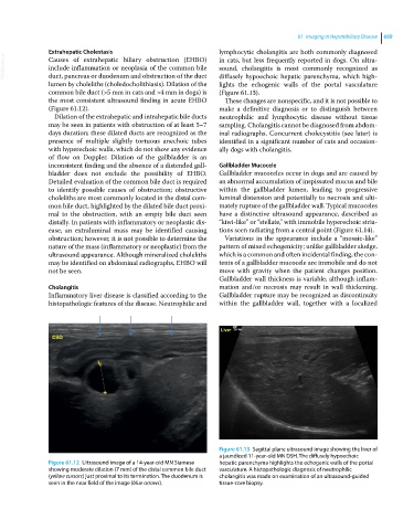
Figure 61.13 Sagittal plane ultrasound image showing the liver of
a jaundiced 11‐year‐old MN DSH. The diffusely hypoechoic
Figure 61.12 Ultrasound image of a 14‐year‐old MN Siamese hepatic parenchyma highlights the echogenic walls of the portal
showing moderate dilation (7 mm) of the distal common bile duct vasculature. A histopathologic diagnosis of neutrophilic
(yellow cursors) just proximal to its termination. The duodenum is cholangitis was made on examination of an ultrasound‐guided
seen in the near field of the image (blue arrows). tissue‐core biopsy.