
Page 705 - Clinical Small Animal Internal Medicine
P. 705
61 Imaging in Hepatobiliary Disease 673
sound; in dogs, the opening of splenorenal collaterals, Larger thrombi can extend from the extrahepatic por-
VetBooks.ir with enlargement of the left gonadal vein and develop- tal vein into the portal branches, resulting in portal
hypertension and acquired shunting.
ment of a complicated plexus of vessels adjacent to the
Radiographic examination of patients with portal
left kidney, has been most commonly documented.
Ultrasound examination may be useful in document- hypertension is often unrewarding. Many will have
ing the cause of portal hypertension. A small, hypere- evidence of ascites, obscuring the serosal detail of the
choic, irregular liver with multiple hypoechoic abdomen. In patients with chronic liver disease, it may
regeneration nodules may be identified in patients with be possible to identify microhepatica, even in the
cirrhosis. Intrahepatic arterioportal fistulae are direct presence of ascites, by the cranial displacement and
connections between the hepatic arteries and the portal clockwise rotation of the gastric axis.
vessels. These vascular anomalies may be congenital or
acquired and may be seen on ultrasound as distended, Peliosis Hepatis
tortuous intrahepatic vessels. The high arterial pressure Peliosis hepatis is seen as randomly distributed cystic and
results in portal hypertension and dramatic ascites, with blood‐filled spaces within the liver, thought to be formed
spectral Doppler demonstrating reversed, often pulsatile due to either focal hepatocytic necrosis or local obstruc-
flow within an enlarged hepatic portal vein. Congenital tion of small portal branches with hepatic atrophy and
hypoplasia of the hepatic portal vein is thought to affect subsequent sinusoidal dilation. The condition is rare in
both extrahepatic and intrahepatic portal vasculature dogs and more commonly identified in older cats. On
and includes the condition of portal vein hypoperfusion ultrasound, the the abnormal sinusoidal dilation may be
previously referred to as microvascular dysplasia. seen as multiple cystic lesions that may mimic vascular
Although hypoplasia of the extrahepatic portal vein tumors, such as hemangioma or hemangiosarcoma.
may be seen as a reduction in vessel diameter, the lack of
objective measurement criteria makes this difficult to
identify definitively. A small liver may be identified in Advanced Imaging Techniques
patients with mild portal vein hypoplasia; ultrasound
examination is often otherwise unremarkable. However, Contrast‐Enhanced Ultrasound
evidence of portal hypertension and acquired portosys-
temic shunting may be identified in patients with more Contrast‐enhanced ultrasound is a non-invasive imag-
severe changes. The most common cause of portal vein ing technique that enables the functional assessment of
obstruction is portal thrombosis, which may occur sec- organ perfusion patterns. The contrast media consist of
ondary to hypercoagulability, vascular stasis or damage microscopic (2–6 μm) inert gas‐filled microbubbles that
to the vascular endothelium (for example, due to local are injected intravenously. When subjected to ultra-
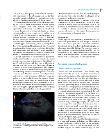
neoplasia or inflammation). Portal thrombosis is recog- sound waves, these bubbles respond with a nonlinear
nized as an intraluminal immobile echogenic structure, oscillation, generating strong harmonic frequencies that
with color Doppler highlighting the thrombus as filling can be detected using specialized transducers and dedi-
defects in the normal pattern of flow (Figure 61.19). cated contrast imaging software. In the liver, two phases
of contrast enhancement may be recognized: the early
phase of enhancement is equivalent to the blood pool
phase and is composed of arterial (wash‐in) and portal
(wash‐out) stages, with the enhancement peaking at the
same time as peak portal blood flow (typically 15–60
seconds in dogs), and disappearing by 150–200 seconds
in most dogs. This early phase allows the detection of
hepatic arteries and small vessels that would not be
apparent with conventional gray‐scale B‐mode imaging.
The late phase is only recognized with contrast media
that are able to leave the blood pool; this phase corre-
sponds to intracellular contrast uptake and enables the
assessment of parenchymal perfusion.
Contrast‐enhanced ultrasound has been demonstrated
as a safe and accurate technique for differentiating
between benign and malignant liver lesions. Malignant
Figure 61.19 Color Doppler ultrasound image highlighting a
portal thrombus as a filling defect within the hepatic portal vein lesions do not have a portal venous supply and are
(blue arrows). perfused only via the hepatic arterial supply. At peak