Page 706 - Clinical Small Animal Internal Medicine
P. 706
674  Section 7  Diseases of the Liver, Gallbladder, and Bile Ducts

            (portal)  early  phase  enhancement,  malignant  nodules   Following the intravenous administration of the radiop-
  VetBooks.ir  therefore appear show hypoenhancement in comparison   harmaceutical   Tc‐mebrofenin, hepatobiliary scintigra-
                                                                          99m
                                                              phy may also used for the evaluation of liver function,
            to the surrounding liver, while benign nodules typically
            show isoenhancement or slight hyperenhancement. A
                                                                                                        99m
                                                              diagnosis of cholecystitis. In the normal patient
            similar pattern is reported during late‐phase enhance-  detection of cholestasis and biliary tract obstruction, and
                                                                                                          Tc‐
            ment, with most malignant lesions appearing relatively   mebrofenin is extracted from the blood pool by the liver,
            hypoechoic.                                       with peak liver radioactivity observed 6–8 minutes after
                                                              injection. The hepatic extraction fraction (HEF) is used as
                                                              a measure of hepatocyte function. Following extraction by
            Nuclear Scintigraphy                              the hepatocytes, the radiopharmaceutical is excreted into
            Although nuclear scintigraphy is a well‐established imag-  the biliary tree with a half‐life of 19 minutes, resulting in
            ing technique, its availability is limited by the strict regula-  detectable radioactivity in the gallbladder and small intes-
            tion of radiopharmaceuticals. Nuclear medicine is most   tines within one hour of injection. A prolonged clearance
            widely used as a very sensitive and minimally invasive   time indicates extrahepatic biliary obstruction and in dogs
            technique for the diagnosis of PSS. The best documented   may allow the detection of abnormalities before any ultra-
            protocol involves the per‐rectal administration of  99m tech-  sound changes are identified.
            netium pertechnetate ( 99m Tc), which is rapidly absorbed
            from the colonic mucosa. The  99m Tc subsequently enters   Computed Tomography
            the mesenteric vessels, from where it should pass through
            the hepatic portal vein into the hepatic parenchyma before   Computed tomography (CT) is an increasingly widely
            filtering through the hepatic microcirculation to enter the   available, rapid and noninvasive diagnostic imaging tech-
            hepatic caudal vena cava and then the heart. In a patient   nique that is especially useful in the evaluation of hepatic
            with a PSS, the  99m Tc bypasses the liver and is delivered   vascular abnormalities  and  hepatic  mass  lesions.
            directly to the heart. Regions of interest (ROIs) drawn   Multidetector row CT angiography enables very detailed
            over the heart and liver are used to calculate the shunt   anatomic evaluation of the hepatic arterial, portal and
            fraction by comparing the intensity of radioactive counts   venous architecture and should facilitate the identifica-
            over the heart with the intensity of counts over the liver.   tion of both intra‐ and extrahepatic portosystemic
                                                              shunts, arterioportal fistulae, and portal vein obstruc-
                                                              tion. Using three-dimensional reconstructions, it should
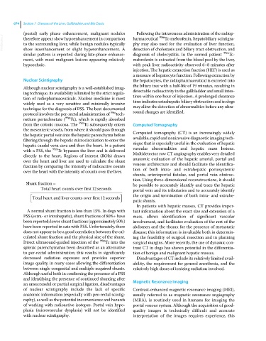
             Shunt fraction                                   be possible to accurately identify and trace the hepatic
                    Total heart counts over first12  seconds    portal vein and its tributaries and to accurately identify
                                                              the  origin and termination of both intra‐ and extrahe-
                Total  heart and liver counts over first12 seconds
                                                              patic shunts.
                                                                In patients with hepatic masses, CT provides impor-
              A normal shunt fraction is less than 15%. In dogs with   tant information about the exact size and extension of a
            PSS (extra‐ or intrahepatic), shunt fractions of 80%+ have   mass, allows identification of significant vascular
            been reported; lower shunt fractions (approximately 50%)   involvement, and facilitates evaluation of the rest of the
            have been reported in cats with PSS. Unfortunately, there   abdomen and the thorax for the presence of metastatic
            does not appear to be a good correlation between the cal-  disease; this information is invaluable both in determin-
            culated shunt fraction and the physical size of the shunt.   ing the feasibility of surgical resection and in planning
            Direct ultrasound‐guided injection of the  99m Tc into the   surgical margins. More recently, the use of dynamic con-
            splenic parenchymahas been described as an alternative   trast CT in dogs has shown potential in the differentia-
            to per‐rectal administration; this results in significantly   tion of benign and malignant hepatic masses.
            decreased radiation exposure and provides superior   Disadvantages of CT include its relatively limited avail-
            image quality, in many cases allowing the differentiation   ability, the requirement for general anesthesia, and the
            between single congenital and multiple acquired shunts.   relatively high doses of ionizing radiation involved.
            Although useful both in confirming the presence of a PSS
            and identifying the presence of continued shunting after
            an unsuccessful or partial surgical ligation, disadvantages   Magnetic Resonance Imaging
            of nuclear scintigraphy include the lack of specific   Contrast‐enhanced magnetic resonance imaging (MRI),
              anatomic information (especially with per‐rectal scintig-  usually referred to as magnetic resonance angiography
            raphy), as well as the potential inconvenience and hazards   (MRA), is routinely used in humans for imaging the
            of working with radioactive isotopes. Portal vein hypo-    portal venous system. Although the acquisition of good‐
            plasia (microvascular dysplasia) will not be identified   quality images is technically difficult and accurate
            with nuclear scintigraphy.                        interpretation of  the images requires experience,  this

