Page 704 - Clinical Small Animal Internal Medicine
P. 704
672  Section 7  Diseases of the Liver, Gallbladder, and Bile Ducts

              Historically, mesenteric portovenography was widely   of the caudal vena cava (between the hepatic veins and
  VetBooks.ir  used for the definitive confirmation of a suspected porto-  right atrium), for example by thrombosis, neoplasia or
                                                              inflammation. Ultrasound findings include hepatomegaly,
            systemic shunt and for the differentiation between intra
            and extrahepatic shunts. The most popular technique for
            portovenography is the intravenous injection of non-  reduced hepatic echogenicity, dilation of the hepatic veins
                                                              and caudal vena cava and, in some cases, the presence of
            ionic iodinated contrast media through a catheter pre-  free abdominal (+/− pleural) fluid. Radiographs may show
            placed into a mesenteric vein. This involves a laparotomy   hepatomegaly and loss of serosal detail in patients with
            and is an invasive procedure. Alternative and potentially   ascites, possibly with evidence of a pleural effusion and
            less invasive, but less commonly used, techniques for   cardiomegaly in patients with right‐sided heart failure.
            contrast administration include percutaneous ultra-
            sound-guided catheterization of the splenic vein, cathe-  Portal Hypertension
            terization of the cranial mesenteric artery via the femoral   Chronic portal hypertension resulting in the develop-
            artery and transvenous retrograde portography. In many   ment of portosystemic collaterals (acquired portosys-
            cases, mesenteric portovenography has been superseded   temic shunts) is relatively common in dogs, but rarely
            by contrast Computed Tomographic examination.     identified in cats. Causes of portal hypertension include
            However, during surgical occlusion of a shunt, portove-  advanced  chronic  liver  disease  (cirrhosis),  intrahepatic
            nography is still used to verify that the correct vessel has   arterioportal fistulae, congenital hypoplasia of the portal
            been occluded, to check that there are no additional   vein and portal vein obstruction. Ultrasound evaluation
            shunting vessels and to assess the degree of portal vascu-  of patients with portal hypertension will typically dem-
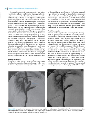
            larization post-ligation. Fluoroscopic imaging of the con-  onstrate ascites, often with edema of the gallbladder wall
            trast injection provides useful real time information (Fig   and pancreas. Spectral Doppler is useful in measuring
            61.18): if this is not available, then standard radiographic   portal velocity; the identification of reduced flow veloc-
            views (ideally both lateral and VD views, each exposed at   ity (from a normal velocity of approximately 17+/-
            the end of a separate contrast injection) should be taken.   5cm/s to <10 cm/s in the dog), and occasionally reversed
                                                              portal flow, is very suggestive of portal hypertension.
            Hepatic Congestion                                The portosystemic collaterals open in response to sus-
            Disturbance of the normal venous outflow results in pas-  tained portal hypertension and connect the portal sys-
            sive hepatic congestion. Causes include right‐sided heart   tem directly to the systemic circulation. These acquired
            failure and obstruction or compression of the cranial part   shunting vessels can be challenging to identify on ultra-


            Figure 61.18  Ventrodorsal intraoperative fluoroscopic image acquired immediately after the administration of nonionic contrast medium
            into a mesenteric vein. The contrast highlights  a left gastro-azygos extrahepatic portosystemic shunt (blue arrow). Source: Rob White,
            School of Veterinary Medicine and Science, University of Nottingham

