
Page 700 - Clinical Small Animal Internal Medicine
P. 700
668 Section 7 Diseases of the Liver, Gallbladder, and Bile Ducts
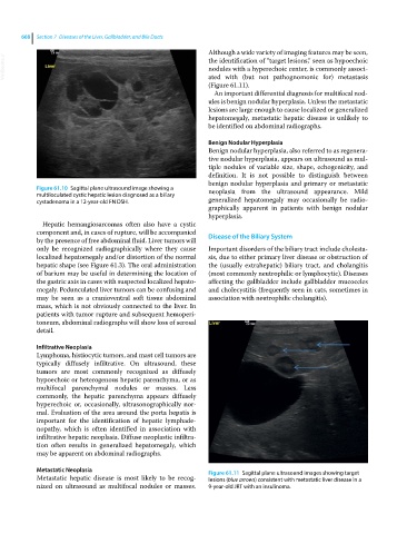
Although a wide variety of imaging features may be seen,
VetBooks.ir the identification of “target lesions,” seen as hypoechoic
nodules with a hyperechoic center, is commonly associ-
ated with (but not pathognomonic for) metastasis
(Figure 61.11).
An important differential diagnosis for multifocal nod-
ules is benign nodular hyperplasia. Unless the metastatic
lesions are large enough to cause localized or generalized
hepatomegaly, metastatic hepatic disease is unlikely to
be identified on abdominal radiographs.
Benign Nodular Hyperplasia
Benign nodular hyperplasia, also referred to as regenera-
tive nodular hyperplasia, appears on ultrasound as mul-
tiple nodules of variable size, shape, echogenicity, and
definition. It is not possible to distinguish between
benign nodular hyperplasia and primary or metastatic
Figure 61.10 Sagittal plane ultrasound image showing a neoplasia from the ultrasound appearance. Mild
multiloculated cystic hepatic lesion diagnosed as a biliary
cystadenoma in a 12‐year‐old FN DSH. generalized hepatomegaly may occasionally be radio-
graphically apparent in patients with benign nodular
hyperplasia.
Hepatic hemangiosarcomas often also have a cystic
component and, in cases of rupture, will be accompanied Disease of the Biliary System
by the presence of free abdominal fluid. Liver tumors will
only be recognized radiographically where they cause Important disorders of the biliary tract include cholesta-
localized hepatomegaly and/or distortion of the normal sis, due to either primary liver disease or obstruction of
hepatic shape (see Figure 61.3). The oral administration the (usually extrahepatic) biliary tract, and cholangitis
of barium may be useful in determining the location of (most commonly neutrophilic or lymphocytic). Diseases
the gastric axis in cases with suspected localized hepato- affecting the gallbladder include gallbladder mucoceles
megaly. Pedunculated liver tumors can be confusing and and cholecystitis (frequently seen in cats, sometimes in
may be seen as a cranioventral soft tissue abdominal association with neutrophilic cholangitis).
mass, which is not obviously connected to the liver. In
patients with tumor rupture and subsequent hemoperi-
toneum, abdominal radiographs will show loss of serosal
detail.
Infiltrative Neoplasia
Lymphoma, histiocytic tumors, and mast cell tumors are
typically diffusely infiltrative. On ultrasound, these
tumors are most commonly recognized as diffusely
hypoechoic or heterogenous hepatic parenchyma, or as
multifocal parenchymal nodules or masses. Less
commonly, the hepatic parenchyma appears diffusely
hyperechoic or, occasionally, ultrasonographically nor-
mal. Evaluation of the area around the porta hepatis is
important for the identification of hepatic lymphade-
nopathy, which is often identified in association with
infiltrative hepatic neoplasia. Diffuse neoplastic infiltra-
tion often results in generalized hepatomegaly, which
may be apparent on abdominal radiographs.
Metastatic Neoplasia Figure 61.11 Sagittal plane ultrasound images showing target
Metastatic hepatic disease is most likely to be recog- lesions (blue arrows) consistent with metastatic liver disease in a
nized on ultrasound as multifocal nodules or masses. 9‐year‐old JRT with an insulinoma.