
Page 697 - Clinical Small Animal Internal Medicine
P. 697
61 Imaging in Hepatobiliary Disease 665
The radiologic assessment of hepatic parenchymal dis- (a)
VetBooks.ir ease is limited to the evaluation of overall hepatic size
and shape; ultrasound assessment provides additional
valuable information about parenchymal echotexture
and echogenicity and is therefore more sensitive in the
detection of parenchymal abnormalities. Hepatic paren-
chymal disease may be described as diffuse, focal or
multifocal, depending on the distribution of changes
identified on ultrasound.
Diffuse Parenchymal Disease
Diffuse parenchymal pathology is usually due to general-
ized infiltrative disease, typically affecting all liver lobes.
Unless the disease process results in a fairly marked
change in liver size and/or shape, the liver may appear
normal on abdominal radiographs. However, in many
cases, changes in hepatic echogenicity and/or echotex-
ture can be identified on ultrasound. (b)
Accurate evaluation of hepatic echogenicity is chal-
lenging, as apparent echogenicity is influenced by
machine settings and transducer choice; this makes the
identification of uniform alterations in hepatic echo-
genicity especially difficult. Comparison of the liver with
adjacent organs imaged at the same depth is useful: the
normal canine liver is coarser than and hypoechoic to
the spleen, but usually isoechoic, or slightly hyperechoic,
to the kidneys. A diffusely hyperechoic liver is likely to
be similar in echogenicity to the spleen, with decreased
contrast between abnormally hyperechoic parenchyma
and the hyperechoic portal vessel walls blurring the nor-
mally clear definition of these walls. A diffusely hypo-
echoic liver may be recognized by the unusual
prominence of the portal vessel walls, which become
highlighted by the abnormally hypoechoic background.
Diffuse parenchymal disease may also be seen as an
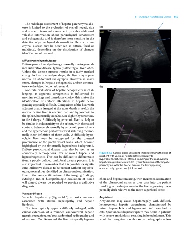
abnormally heterogenous liver of mixed hyper‐ and Figure 61.6 Sagittal plane ultrasound images showing the liver of
hypoechogenicity. This can be difficult to differentiate a patient with vacuolar hepatopathy secondary to
from a poorly defined multifocal disease process. It is hyperadrenocorticism. (a) Marked rounding of the caudoventral
hepatic margin (blue arrows). (b) Hyperattenuation of the hepatic
also important to remember that it is possible for signifi- parenchyma, with the deeper areas of the liver appearing
cant infiltrative disease to be present without any obvi- unexpectedly hypoechoic (pink arrows).
ous abnormalities identified on ultrasound examination.
Due to the nonspecific nature of the imaging findings,
cytologic and/or histopathologic evaluation of tissue choic and hyperattenuating, with increased attenuation
will almost always be required to provide a definitive of the ultrasound waves as they pass into the patient
diagnosis. resulting in the deeper areas of the liver appearing unex-
pectedly dark relative to the more superficial areas.
Vacuolar Disease
Vacuolar hepatopathy (Figure 61.6) is most commonly Amyloidosis
associated with steroid hepatopathy and hepatic Amyloidosis may cause hepatomegaly, with diffusely
lipidosis. heterogenous hepatic parenchyma characterized by
The liver typically appears diffusely enlarged, with mixed hyperechoic and hypoechoic foci described in
caudal extension of a rounded caudoventral hepatic cats. Spontaneous hepatic rupture may occur in patients
margin recognized on both abdominal radiography and with severe amyloidosis, resulting in hemabdomen. This
ultrasound. On ultrasound, the liver is typically hypere- would be recognized on abdominal radiographs as loss