
Page 698 - Clinical Small Animal Internal Medicine
P. 698
666 Section 7 Diseases of the Liver, Gallbladder, and Bile Ducts
of normal serosal detail and on ultrasound as the pres- Hepatocutaneous Syndrome
VetBooks.ir ence of free abdominal fluid. A breed predisposition to superficial necrolytic dermatitis, is occasionally seen in
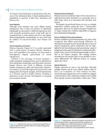
Hepatocutaneous syndrome (Figure 61.8), also known as
amyloidosis is reported in Shar Peis, Abyssinian and
older dogs, where it is associated with ulcerative skin
Siamese cats.
lesions.
Acute Hepatitis The hepatic parenchymal lesions are seen as multiple
Although acute hepatitis may cause diffuse hepatic hypoechoic nodules, giving the liver a “honeycomb”
enlargement, this is often not obvious on abdominal appearance. There are no marked changes in hepatic size
radiography or ultrasound. A diffusely hypoechoic liver or shape, making this condition impossible to diagnose
with unusually prominent portal vessel walls may be from abdominal radiographs.
recognized but in some patients the liver may appear
ultrasonographically unremarkable. In cats, there may Focal or Multifocal Parenchymal Disease
be evidence of concurrent biliary tract disease (see Focal or multifocal parenchymal lesions are more easily
later). recognized than diffuse parenchymal infiltration on
ultrasound examination and are typically seen as areas of
Chronic Hepatitis and Cirrhosis altered echogenicity and/or echotexture that are high-
Chronic hepatitis (Figure 61.7) is usually associated lighted by the surrounding normal parenchyma. Large or
with a mixed pattern of hepatic fibrosis, inflamma- peripheral lesions may be identified on abdominal radio-
tion, and hyperplasia; this appears on ultrasound as graphs if they result in localized hepatomegaly or distor-
heterogenous hepatic parenchyma of mixed tion of the hepatic margins. The imaging features
echogenicity. described are not specific and it is not possible to accu-
Cirrhosis is the end‐stage of chronic hepatitis; a rately differentiate the different lesions on imaging
small, irregularly marginated liver may be identified on appearance alone.
both abdominal radiography and ultrasound; extensive
hepatic fibrosis may result in diffusely increased Hepatic Abscesses
hepatic echogenicity, often with hypoechoic regenera- Hepatic abscesses are rare in cats and dogs. Although
tive nodules scattered throughout the parenchyma and multiple lesions may be seen, solitary lesions, often at
bulging from the hepatic margins. Portal hypertension least 3 cm in diameter, are more common. On ultra-
is a common sequel to hepatic fibrosis, resulting in sound, the typical appearance is of a rounded or irregular
abdominal ascites and the development of acquired cavitated lesion, with a relatively echogenic and variably
portosystemic shunts. thick capsule around a hypoechoic central area. There
Figure 61.8 Sagittal plane ultrasound image showing the liver of
a Cavalier King Charles spaniel presented with ulcerative skin
Figure 61.7 Sagittal plane ultrasound image showing the liver of lesions. Multiple poorly defined hypoechoic nodules are seen
a patient with chronic hepatitis. The liver is small and irregularly throughout the hepatic parenchyma (blue arrows);
marginated (blue arrows). hepatocutaneous syndrome was diagnosed from a liver biopsy.