
Page 702 - Clinical Small Animal Internal Medicine
P. 702
670 Section 7 Diseases of the Liver, Gallbladder, and Bile Ducts
VetBooks.ir
Figure 61.14 Ultrasound image showing the typical “kiwi‐like”
appearance of a gallbladder mucocele in a 5‐year‐old FN
Chihuahua.
collection of free fluid and abnormally hyperechoic fat
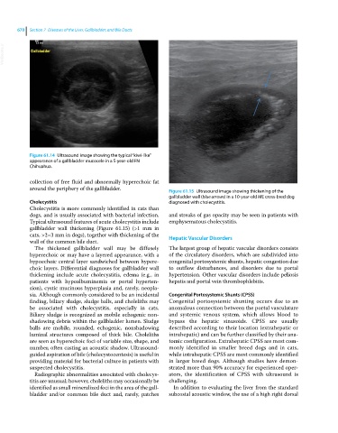
around the periphery of the gallbladder. Figure 61.15 Ultrasound image showing thickening of the
gallbladder wall (blue arrows) in a 10‐year‐old ME cross‐bred dog
Cholecystitis diagnosed with cholecystitis.
Cholecystitis is more commonly identified in cats than
dogs, and is usually associated with bacterial infection. and streaks of gas opacity may be seen in patients with
Typical ultrasound features of acute cholecystitis include emphysematous cholecystitis.
gallbladder wall thickening (Figure 61.15) (>1 mm in
cats, >2–3 mm in dogs), together with thickening of the Hepatic Vascular Disorders
wall of the common bile duct.
The thickened gallbladder wall may be diffusely The largest group of hepatic vascular disorders consists
hyperechoic or may have a layered appearance, with a of the circulatory disorders, which are subdivided into
hypoechoic central layer sandwiched between hypere- congenital portosystemic shunts, hepatic congestion due
choic layers. Differential diagnoses for gallbladder wall to outflow disturbances, and disorders due to portal
thickening include acute cholecystitis, edema (e.g., in hypertension. Other vascular disorders include peliosis
patients with hypoalbuminemia or portal hyperten- hepatis and portal vein thrombophlebitis.
sion), cystic mucinous hyperplasia and, rarely, neopla-
sia. Although commonly considered to be an incidental Congenital Portosystemic Shunts (CPSS)
finding, biliary sludge, sludge balls, and choleliths may Congenital portosystemic shunting occurs due to an
be associated with cholecystitis, especially in cats. anomalous connection between the portal vasculature
Biliary sludge is recognized as mobile echogenic non- and systemic venous system, which allows blood to
shadowing debris within the gallbladder lumen. Sludge bypass the hepatic sinusoids. CPSS are usually
balls are mobile, rounded, echogenic, nonshadowing described according to their location (extrahepatic or
luminal structures composed of thick bile. Choleliths intrahepatic) and can be further classified by their ana-
are seen as hyperechoic foci of variable size, shape, and tomic configuration. Extrahepatic CPSS are most com-
number, often casting an acoustic shadow. Ultrasound‐ monly identified in smaller breed dogs and in cats,
guided aspiration of bile (cholecystocentesis) is useful in while intrahepatic CPSS are most commonly identified
providing material for bacterial culture in patients with in larger breed dogs. Although studies have demon-
suspected cholecystitis. strated more than 90% accuracy for experienced oper-
Radiographic abnormalities associated with cholecys- ators, the identification of CPSS with ultrasound is
titis are unusual; however, choleliths may occasionally be challenging.
identified as small mineralized foci in the area of the gall- In addition to evaluating the liver from the standard
bladder and/or common bile duct and, rarely, patches subcostal acoustic window, the use of a high right dorsal