
Page 703 - Clinical Small Animal Internal Medicine
P. 703
61 Imaging in Hepatobiliary Disease 671
acoustic window, located just cranial to the right kidney, Comparison of the diameter of the portal vein at the
VetBooks.ir with the patient positioned in left lateral recumbency, is porta hepatis with the diameter of the aorta and caudal
vena cava at the same level is very useful; a portal
recommended for visualization of the caudal vena cava
(CVC) and the hepatic portal vein (HPV) as they enter
extrahepatic shunt. Conversely, an extrahepatic shunt is
the liver; in most patients with an extrahepatic CPSS, vein:aortic ratio ≤0.65 is predictive for the presence of an
this window allows identification of the shunting vessel considered unlikely with a portal vein:aortic ratio >0.8
itself. Subcostal, right dorsal intercostal, and left ventral and a portal vein:caval ratio >0.75.
intercostal windows are all recommended for evaluation Intrahepatic CPSS may be classified as left, central or
of the liver in patients with a suspected intrahepatic right divisional. The left divisional shunt, which repre-
CPSS. In anesthetized patients with a very small liver, sents a patent ductus venosus and joins the left portal
positive pressure ventilation may be useful in improving branch to the intrahepatic caudal vena cava, is the most
visualization by displacing the liver caudally. Although common (Figure 61.17).
not essential, color and spectral Doppler allow the deter- Both right and central divisional shunts arise from the
mination of flow direction, the identification of turbu- right portal branch; right divisional shunts are long
lence and the measurement of hepatic portal velocity, shunts, whereas the central divisional shunt is usually a
and are therefore very useful in the ultrasound assess- short aneurysm‐like dilation joining the right portal
ment of patients with suspected CPSS. branch with the intrahepatic caudal vena cava.
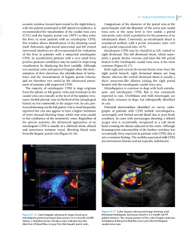
The majority of extrahepatic CPSS in dogs originate Microhepatica is common in dogs with both extrahe-
from the splenic or left gastric veins and terminate in the patic and intrahepatic CPSS, but is less commonly
caudal vena cava (usually at the level of the epiploic fora- reported in cats. Urolithiasis and mild renomegaly are
men), the left phrenic vein (at the level of the oesophageal also fairly common in dogs, but infrequently identified
hiatus) or, less commonly, in the azygos vein. In cats, por- in cats.
tocaval shunting via the left gastric vein is most frequently Potential abnormalities identified on survey radio-
reported but cats also appear to have a higher incidence graphs of patients with CPSS include microhepatica,
of more unusual shunting loops, which may arise caudal renomegaly, and limited serosal detail due to poor body
to the confluence of the mesenteric veins. Regardless of condition. In cases with portoazygos shunting, a dilated
the precise anatomy, the ultrasound appearance of an azygos vein is occasionally recognized as a soft tissue
extrahepatic CPSS is usually of a relatively short, dilated band crossing the thorax adjacent to the aorta. Although
and sometimes tortuous vessel, diverting blood away hematogenous osteomyelitis of the lumbar vertebrae has
from the hepatic portal vein (Figure 61.16). occasionally been reported in patients with CPSS, this is
not a common finding. The uroliths associated with CPSS
are ammonium biurate and are typically radiolucent.
Figure 61.17 Color Doppler ultrasound image showing a left
Figure 61.16 Color Doppler ultrasound image showing an divisional intrahepatic portocaval shunt in a 6‐month‐old FE
extrahepatic portocaval shunt (blue arrows) in a 6‐month‐old ME golden retriever. The mosaic pattern of the color Doppler indicates
Bichon × Yorkshire terrier. The blue color indicates that the turbulence at the point that the shunt joins the intrahepatic
direction of blood flow is away from the hepatic portal vein. caudal vena cava.