
Page 119 - Feline diagnostic imaging
P. 119
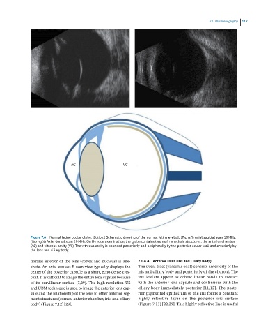
7.1 ltrasonograahy 117
AC VC
Figure 7.6 Normal feline ocular globe. (Bottom) Schematic drawing of the normal feline eyeball. (Top left) Axial sagittal scan 10 MHz.
(Toa right) Axial dorsal scan 10 MHz. On B-mode examination, the globe contains two main anechoic structures: the anterior chamber
(AC) and vitreous cavity (VC). The vitreous cavity is bounded posteriorly and peripherally by the posterior ocular wall and anteriorly by
the lens and ciliary body.
normal interior of the lens (cortex and nucleus) is ane- 7.1.4.4 Anterior Uvea (Iris and Ciliary Body)
choic. An axial contact B‐scan view typically displays the The uveal tract (vascular coat) consists anteriorly of the
center of the posterior capsule as a short, echo‐dense cres- iris and ciliary body and posteriorly of the choroid. The
cent. It is difficult to image the entire lens capsule because iris leaflets appear as echoic linear bands in contact
of its curvilinear surface [7,29]. The high‐resolution US with the anterior lens capsule and continuous with the
and UBM technique is used to image the anterior lens cap- ciliary body immediately posterior [11,12]. The poste -
sule and the relationship of the lens to other anterior seg- rior pigmented epithelium of the iris forms a constant
ment structures (cornea, anterior chamber, iris, and ciliary highly reflective layer on the posterior iris surface
body) (Figure 7.12) [29]. (Figure 7.13) [22,29]. This highly reflective line is useful