
Page 120 - Feline diagnostic imaging
P. 120
118 7 Normal Cross-sectional Anatomy of the Eye and Orbit
C A P R S
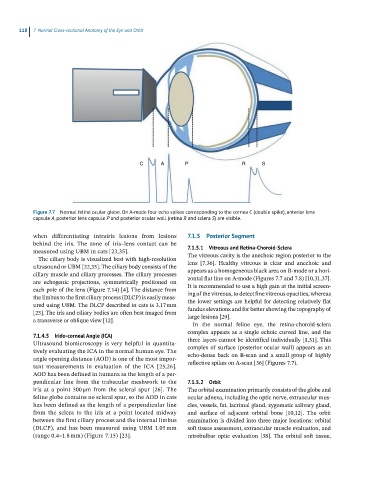
Figure 7.7 Normal feline ocular globe. On A-mode four echo spikes corresponding to the cornea C (double spike), anterior lens
capsule A, posterior lens capsule P and posterior ocular wall (retina R and sclera S) are visible.
when differentiating intrairis lesions from lesions 7.1.5 Posterior Segment
behind the iris. The zone of iris–lens contact can be
measured using UBM in cats [23,35]. 7.1.5.1 Vitreous and Retina-Choroid-Sclera
The vitreous cavity is the anechoic region posterior to the
The ciliary body is visualized best with high‐resolution
ultrasound or UBM [22,35]. The ciliary body consists of the lens [7,36]. Healthy vitreous is clear and anechoic and
appears as a homogeneous black area on B‐mode or a hori-
ciliary muscle and ciliary processes. The ciliary processes
are echogenic projections, symmetrically positioned on zontal flat line on A‐mode (Figures 7.7 and 7.8) [10,31,37].
It is recommended to use a high gain at the initial screen-
each pole of the lens (Figure 7.14) [4]. The distance from
the limbus to the first ciliary process (DLCP) is easily meas- ing of the vitreous, to detect fine vitreous opacities, whereas
the lower settings are helpful for detecting relatively flat
ured using UBM. The DLCP described in cats is 3.17 mm
[23]. The iris and ciliary bodies are often best imaged from fundus elevations and for better showing the topography of
large lesions [29].
a transverse or oblique view [12].
In the normal feline eye, the retina‐choroid‐sclera
complex appears as a single echoic curved line, and the
7.1.4.5 Irido-corneal Angle (ICA)
Ultrasound biomicroscopy is very helpful in quantita- three layers cannot be identified individually [1,31]. This
complex of surface (posterior ocular wall) appears as an
tively evaluating the ICA in the normal human eye. The
angle opening distance (AOD) is one of the most impor - echo‐dense back on B‐scan and a small group of highly
reflective spikes on A‐scan [36] (Figures 7.7).
tant measurements in evaluation of the ICA [25,26].
AOD has been defined in humans as the length of a per -
pendicular line from the trabecular meshwork to the 7.1.5.2 Orbit
iris at a point 500 μm from the scleral spur [26]. The The orbital examination primarily consists of the globe and
feline globe contains no scleral spur, so the AOD in cats ocular adnexa, including the optic nerve, extraocular mus-
has been defined as the length of a perpendicular line cles, vessels, fat, lacrimal gland, zygomatic salivary gland,
from the sclera to the iris at a point located midway and surface of adjacent orbital bone [10,12]. The orbit
between the first ciliary process and the internal limbus examination is divided into three major locations: orbital
(DLCP), and has been measured using UBM 1.05 mm soft tissue assessment, extraocular muscle evaluation, and
(range 0.4–1.8 mm) (Figure 7.15) [23]. retrobulbar optic evaluation [38]. The orbital soft tissue,