
Page 125 - Feline diagnostic imaging
P. 125
7.2 Comauted Tomograahy and agnetic Resonance maging 123
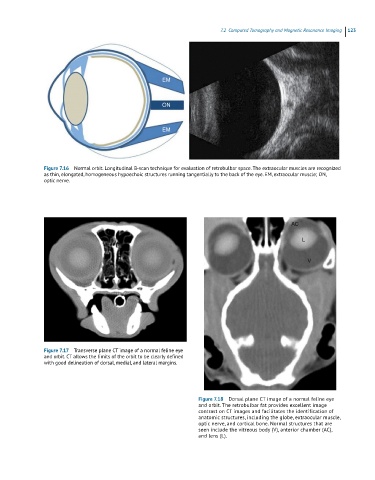
EM
ON
EM
Figure 7.16 Normal orbit. Longitudinal B-scan technique for evaluation of retrobulbar space. The extraocular muscles are recognized
as thin, elongated, homogeneous hypoechoic structures running tangentially to the back of the eye. EM, extraocular muscle; ON,
optic nerve.
Figure 7.17 Transverse plane CT image of a normal feline eye
and orbit. CT allows the limits of the orbit to be clearly defined
with good delineation of dorsal, medial, and lateral margins.
Figure 7.18 Dorsal plane CT image of a normal feline eye
and orbit. The retrobulbar fat provides excellent image
contrast on CT images and facilitates the identification of
anatomic structures, including the globe, extraocular muscle,
optic nerve, and cortical bone. Normal structures that are
seen include the vitreous body (V), anterior chamber (AC),
and lens (L).