
Page 126 - Feline diagnostic imaging
P. 126
124 7 Normal Cross-sectional Anatomy of the Eye and Orbit
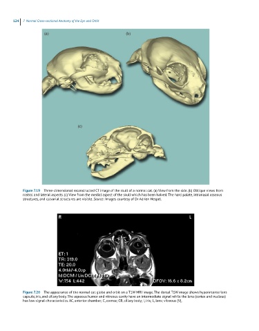
Figure 7.19 Three-dimensional reconstructed CT image of the skull of a normal cat. (a) View from the side. (b) Oblique views from
rostral and lateral aspects. (c) View from the medial aspect of the skull which has been halved. The hard palate, intranasal osseous
structures, and calvarial structures are visible. Source: Images courtesy of Dr Adrien Hespel.
Figure 7.20 The appearance of the normal cat globe and orbit on a T1W MRI image. The dorsal T1W image shows hyperintense lens
capsule, iris, and ciliary body. The aqueous humor and vitreous cavity have an intermediate signal while the lens (cortex and nucleus)
has low signal characteristics. AC, anterior chamber; C, cornea; CB, ciliary body; I, iris; L, lens; vitreous (V),