
Page 121 - Feline diagnostic imaging
P. 121
7.1 ltrasonograahy 119
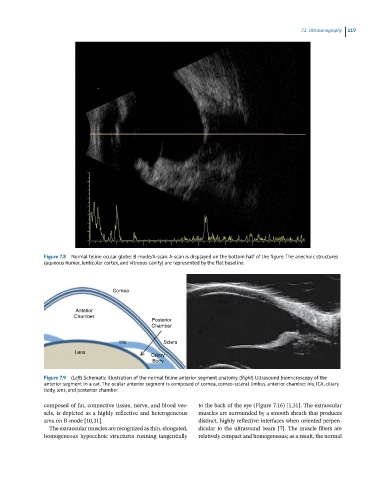
Figure 7.8 Normal feline ocular globe: B-mode/A-scan. A-scan is displayed on the bottom half of the figure. The anechoic structures
(aqueous humor, lenticular cortex, and vitreous cavity) are represented by the flat baseline.
Cornea
Anterior
Chamber
Posterior
Chamber
Iris Sclera
Lens
Ciliary
Body
Figure 7.9 (Left) Schematic illustration of the normal feline anterior segment anatomy. (Right) Ultrasound biomicroscopy of the
anterior segment in a cat. The ocular anterior segment is composed of cornea, corneo-scleral limbus, anterior chamber, iris, ICA, ciliary
body, lens, and posterior chamber.
composed of fat, connective tissue, nerve, and blood ves- to the back of the eye (Figure 7.16) [1,31]. The extraocular
sels, is depicted as a highly reflective and heterogeneous muscles are surrounded by a smooth sheath that produces
area on B‐mode [10,31]. distinct, highly reflective interfaces when oriented perpen-
The extraocular muscles are recognized as thin, elongated, dicular to the ultrasound beam [7]. The muscle fibers are
homogeneous hypoechoic structures running tangentially relatively compact and homogeneous; as a result, the normal