
Page 373 - Feline diagnostic imaging
P. 373
382 23 Liver
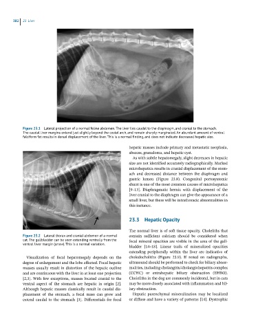
Figure 23.1 Lateral projection of a normal feline abdomen. The liver lies caudal to the diaphragm, and cranial to the stomach.
The caudal liver margins extend just slightly beyond the costal arch, and remain sharply marginated. An abundant amount of ventral
falciform fat results in dorsal displacement of the liver. This is a normal finding, and does not indicate decreased hepatic size.
hepatic masses include primary and metastatic neoplasia,
abscess, granuloma, and hepatic cyst.
As with subtle hepatomegaly, slight decreases in hepatic
size are not identified accurately radiographically. Marked
microhepatica results in cranial displacement of the stom-
ach and decreased distance between the diaphragm and
gastric lumen (Figure 23.8). Congenital portosystemic
shunt is one of the most common causes of microhepatica
[9–13]. Diaphragmatic hernia with displacement of the
liver cranial to the diaphragm can give the appearance of a
small liver, but there will be intrathoracic abnormalities in
this instance.
23.3 Hepatic Opacity
The normal liver is of soft tissue opacity. Choleliths that
Figure 23.2 Lateral thorax and cranial abdomen of a normal contain sufficient calcium should be considered when
cat. The gallbladder can be seen extending ventrally from the focal mineral opacities are visible in the area of the gall-
ventral liver margin (arrow). This is a normal variation.
bladder [14–18]. Linear trails of mineralized opacities
extending peripherally within the liver are indicative of
Visualization of focal hepatomegaly depends on the choledocholiths (Figure 23.9). If noted on radiographs,
degree of enlargement and the lobe affected. Focal hepatic ultrasound should be performed to check for biliary abnor-
masses usually result in distortion of the hepatic outline malities, including cholangitis/cholangiohepatitis complex
and are continuous with the liver in at least one projection (CCHC) or extrahepatic biliary obstruction (EHBO).
[2,3]. With few exceptions, masses located cranial to the Choleliths in the dog are commonly incidental, but in cats
ventral aspect of the stomach are hepatic in origin [2]. may be more closely associated with inflammation and bil-
Although hepatic masses classically result in caudal dis- iary obstruction.
placement of the stomach, a focal mass can grow and Hepatic parenchymal mineralization may be localized
extend caudal to the stomach [3]. Differentials for focal or diffuse and have a variety of patterns [14]. Dystrophic