
Page 378 - Feline diagnostic imaging
P. 378
23.5 epatic Ultrasound 387
23.4 Contrast Radiography:
Portography
Portosystemic shunts are congenital or acquired anomalies
with an abnormal communication between the portal and
systemic venous systems. The anomalous shunting vessel
diverts portal blood into a systemic vein (caudal vena
cava, azygous vein most commonly), bypassing the liver.
Extrahepatic shunts, typically left gastric vein to caudal
vena cava, are the most common congenital shunts in the
cat [9,12,25–28]. Congenital intrahepatic shunts are less
common, with left divisional (patent ductus venosus) seen
most frequently [9,12,25–28]. Various imaging techniques
have been used to demonstrate the anomalous vessels,
including cranial mesenteric portography, percutaneous
splenoportography, and operative mesenteric portography
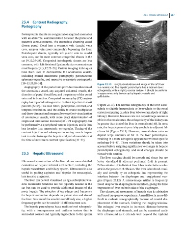
[10–12,25,28–31]. Figure 23.10 Longitudinal ultrasound image of the left liver
Angiography of the portal vein provides visualization of in a normal cat. The hepatic parenchyma has a medium level
the anomalous vessel, any acquired collateral vessels, the echogenicity, with a slightly coarse texture. It should be uniform
direction of portal blood flow, and the patency of the portal in appearance, only broken up by hepatic vessels and
gallbladder.
vein and its branches. Computed tomography (CT) angiog-
raphy has replaced intraoperative contrast injection in most
patients [32,33]. Fast scan times, good spatial, contrast, and (Figure 23.10). The normal echogenicity of the liver is iso-
temporal resolution, and the ability to create multiplanar echoic to slightly hyperechoic or hypoechoic to the renal
and three‐dimensional images allow excellent visualization cortex (comparing caudate liver lobe to cranial pole of right
of anomalous vessels, with more exact determination of kidney). However, because cats can deposit large amounts
origin and termination locations [34]. CT angiography can of fat in the renal cortex, the echogenicity of the kidney can
be performed via a peripheral venous injection, and is thus be greater than that of the liver in normal cats [40]. In most
less invasive than mesenteric portography. Timing of the cats, the hepatic parenchyma is hypoechoic to adjacent fal-
contrast injection and subsequent scanning runs is impor- ciform fat (Figure 23.11). However, normal obese cats can
tant in order to image the hepatic and portal vasculature at deposit large amounts of fat in the liver parenchyma,
the time of maximum contrast opacification [33–39]. resulting in a more echogenic appearance without specific
pathology [41–43]. These variations should be taken into
account before assigning significance to changes in hepatic
parenchymal echogenicity, and mild changes should be
23.5 Hepatic Ultrasound viewed with caution.
The liver margins should be smooth and sharp but are
Ultrasound examination of the liver allows more detailed better visualized if adjacent peritoneal fluid is present.
evaluation of hepatic internal architecture, including the Differentiation of individual liver lobes is also better evalu-
hepatic vasculature and biliary system. Ultrasound is also ated in the presence of effusion. The liver is bordered crani-
useful in guiding aspirates and biopsies for nonsurgical, ally and dorsally by an echogenic line representing the
less invasive diagnoses. interface between the diaphragm and lung/pleural mar-
The liver can be well visualized using a subxiphoid win- gins (Figure 23.12). A mirror‐image artifact is frequently
dow. Intercostal windows are not typically needed in the noted deep to the diaphragmatic interface, giving the false
cat but can be used to provide additional images of the impression of liver on both sides of the diaphragm.
porta hepatis. The selection of transducer and frequency The ultrasound assessment of hepatic size is subjective
for hepatic evaluation depends on patient size and size of and based on operator experience. A small liver is more dif-
the liver. Because of the smaller overall body size, a higher ficult to evaluate sonographically because of cranial dis-
frequency probe can be used (8–12 MHz) in most cats. placement of the stomach, limiting the imaging window.
The hepatic parenchyma has a medium‐level echogenic- The enlarged liver results in increased distance between
ity, with a homogeneous and uniform texture that is the diaphragm and stomach, and can be examined easily
somewhat coarser and typically hypoechoic to the spleen with ultrasound as it extends well beyond the xiphoid