
Page 379 - Feline diagnostic imaging
P. 379
388 23 Liver
(a) (b)
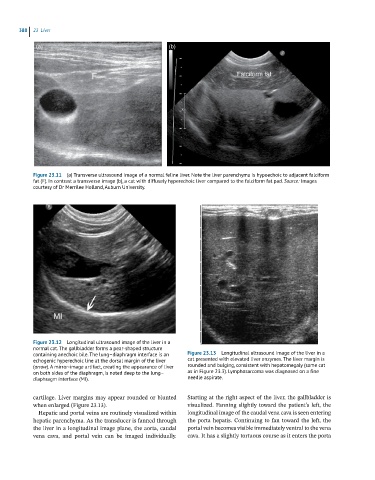
Figure 23.11 (a) Transverse ultrasound image of a normal feline liver. Note the liver parenchyma is hypoechoic to adjacent falciform
fat (F). In contrast a transverse image (b), a cat with diffusely hyperechoic liver compared to the falciform fat pad. Source: Images
courtesy of Dr Merrilee Holland, Auburn University.
Figure 23.12 Longitudinal ultrasound image of the liver in a
normal cat. The gallbladder forms a pear-shaped structure
containing anechoic bile. The lung–diaphragm interface is an Figure 23.13 Longitudinal ultrasound image of the liver in a
echogenic hyperechoic line at the dorsal margin of the liver cat presented with elevated liver enzymes. The liver margin is
(arrow). A mirror-image artifact, creating the appearance of liver rounded and bulging, consistent with hepatomegaly (same cat
on both sides of the diaphragm, is noted deep to the lung– as in Figure 23.3). Lymphosarcoma was diagnosed on a fine
diaphragm interface (MI). needle aspirate.
cartilage. Liver margins may appear rounded or blunted Starting at the right aspect of the liver, the gallbladder is
when enlarged (Figure 23.13). visualized. Fanning slightly toward the patient’s left, the
Hepatic and portal veins are routinely visualized within longitudinal image of the caudal vena cava is seen entering
hepatic parenchyma. As the transducer is fanned through the porta hepatis. Continuing to fan toward the left, the
the liver in a longitudinal image plane, the aorta, caudal portal vein becomes visible immediately ventral to the vena
vena cava, and portal vein can be imaged individually. cava. It has a slightly tortuous course as it enters the porta